-
Phone
13145925686
-
Address
6th Floor, Building 2, Honghui Technology Park, Liuxian 2nd Road, Xin'an Street, Bao'an District, Shenzhen City, Guangdong Province
Product Categories
- Spark direct reading spectrometer
- Rheometer
- TGA
- Chemical pretreatment
- Wavelength dispersive X-ray fluorescence spectrometer
- Differential thermal scanner
- Precious metal detection
- Optics and Scanning Electron Microscopy
- Material film thickness gauge
- Inductively Coupled Plasma Emission Spectrometer
- Oil testing
- Jewelry testing
- Standard Sample
- Glow discharge emission spectrometer
- Nano particle size analyzer
- Physical Materials Laboratory Supporting Equipment
- scanning electron microscope
- Energy Dispersive X-Ray Fluoresence Spectrometer
Shenzhen Huapu General Technology Co., Ltd
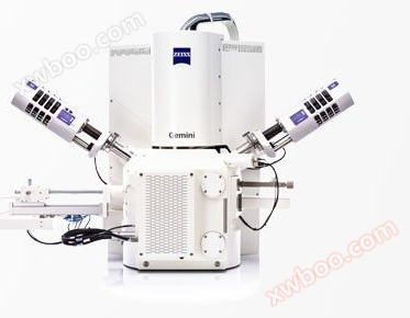
German Zeiss Field Emission Scanning Electron Microscope Sigma Series Products - Huapu General
NegotiableUpdate on 06/19
- Model
- Nature of the Manufacturer
- Producers
- Product Category
- Place of Origin
Overview
German Zeiss Sigma series products for field emission scanning electron microscopy for high-quality imaging and advanced analysis
Product Details
Flexible detection, 4-step workflow, advanced analytical performance
Combining advanced analytical performance with field emission scanning technology, utilizing mature Gemini electronic optical components. Multiple detectors to choose from: for imaging particles, surfaces, or nanostructures. Sigma's semi-automatic 4-step workflow saves a lot of time: setting up imaging and analysis steps to improve efficiency.

Flexible detection for clear imaging
Customize Sigma for your needs using advanced detection techniques to characterize all samples.
Utilize in lens dual detectors to obtain morphology and composition information.
By utilizing the new generation of secondary detectors, up to 50% of the signal images can be obtained. Utilizing Sigma's innovative C2D and variable pressure detector in variable pressure mode, sharp images with up to 85% contrast can be obtained in low vacuum environments.

Automation accelerates workflow
The 4-step workflow allows you to control all functions of Sigma. In a multi-user environment, to achieve fast imaging and save training, first navigate the sample and then set imaging conditions.
Firstly, navigate the sample and then set the imaging conditions.
Next, optimize the areas of interest in the sample and automatically capture images. Finally, use the final step of the workflow to visualize the results.

Advanced analytical microscope
Combining scanning electron microscopy with fundamental analysis: Sigma's top-notch backscattered geometry detector greatly enhances analytical performance, especially for samples sensitive to electron beams.
Obtain analysis data under half of the detection beam and twice the speed conditions.
Benefiting from a short analysis working distance of 8.5 mm and a 35 ° angle, complete and shadow free analysis results were obtained.

Based on mature Gemini technology
The design of Gemini lenses takes into account the influence of electric and magnetic fields on optical performance, and reduces the impact of the field on the sample to a lower level. This enables excellent imaging results even for magnetic samples.
The detection of Gemini in lens ensures the efficiency of signal detection, reducing imaging time simultaneously through secondary detection (SE) and backscatter (BSE) elements.
The Gemini electron beam accelerator technology ensures small detector size and high signal-to-noise ratio.
Flexible detection for clear imaging
Use new detection techniques to characterize all samples.
Utilizing innovative ETSE and in lens detectors in high vacuum mode to obtain morphology and high-resolution information.
Obtain sharp images using variable pressure secondary electrons and C2D detectors in variable pressure mode.
Generate high-resolution transmission images using aSTEM detectors.
Perform component analysis using BSD or YAG detectors.
accessory

SmartEDX
Bringing you an integrated energy spectrum analysis solution
If SEM imaging technology alone cannot fully understand the components or samples, researchers need to use energy dispersive spectroscopy (EDS) in SEM for microscopic analysis. By optimizing the energy spectrum solution for low voltage applications, you can obtain spatial distribution information of elemental chemical composition. Thanks to:
Optimized conventional microscopic analysis applications, and due to the excellent transmittance of silicon nitride windows, it can detect low-energy X-rays of light elements.
The workflow guided graphical user interface greatly improves usability and repeatability in multi-user environments.
Complete service and system support, with Zeiss engineers providing one-stop service for your installation, preventive maintenance, and warranty.

Raman imaging combined with scanning electron microscopy system
Fully integrated Raman imaging
Add Raman spectroscopy and imaging results to your data to obtain richer characterization information of the material. By expanding the Zeiss Sigma 300 with confocal Raman imaging capabilities, you can obtain unique chemical fingerprint information in the sample to identify its composition.
Identify molecular and crystal structure information
3D analysis can be performed, and SEM images, Raman surface scan imaging, and EDS data can be correlated when needed.
Fully integrated RISE allows you to experience the advantages brought by advanced SEM and Raman systems.
Similar Product Recommend


