-
Phone
1376488515213764885152
-
Address
Building 6, No. 1150 Lanfeng Road, Fengxian District, 4597
Product Categories
Shanghai Jintong Instrument Co., Ltd

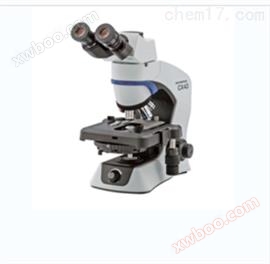
BX63 Olympus fully intelligent electric microscope BX63 parameter quotation picture
NegotiableUpdate on 03/19
- Model
- Nature of the Manufacturer
- Producers
- Product Category
- Place of Origin
Overview
Olympus fully intelligent electric microscope BX63 parameter quotation picture * Electrified, and achieved * focusing through objective lens converter instead of stage, so as to fix the stage and make it more stable. The electrified stage also has new features, driven by advanced high-precision ultrasonic Piezo technology, which can achieve quiet, smooth, and quite precise operation. The stage can be positioned by hand, allowing for quick alignment of large specimens and continuous reading of X and Y positions. Excellent decoding enables users to set precise coordinates and move directly to that location at high speed.
Product Details
Olympus fully intelligent electric microscope BX63 parameter quotation picture
Olympus fully intelligent electric microscope BX63 parameter quotation pictureShanghai Jintong Instrument provides nationwide quotation data
*Electrification and focusing can be achieved through an objective lens converter instead of a stage, which can fix the stage and make it more stable. The electrified stage also has new features, driven by advanced high-precision ultrasonic Piezo technology, which can achieve quiet, smooth, and quite precise operation. The stage can be positioned by hand, allowing for quick alignment of large specimens, while highly accurate decoding enables continuous reading of X and Y positions. Excellent decoding enables users to set precise coordinates and move directly to that location at high speed.
Intelligent design, both flexible and reliable
BX63 is in response to many researchers andmicroscopeAn electric microscope developed based on user requirements. With just a simple touch, the microscope can be quickly operated through the detachable, freely positioned U-MCZ controller or a computer controlled by cellSens imaging software.
Using cellSens software to feedback all data collected by controlling the microscope through a touch screen or U-MCZ controller, allowing users to freely operate the microscope in a more convenient way. From image capture to report generation, Olympus cellSens software personalizes each researcher's operating environment based on their workflow.
* cellSensThe software is not used for clinical diagnosis.

Stability for improving imaging reliability
BX63 utilizes an electrically powered Z-axis objective converter (possibly thanks to infinity correction optics), as well asstageFixed to the idle area of the frame at two additional points for focusing, making it quite stable.

Accurate electric focusing and field of view aperture
The advantage of electrification of the objective lens converter is that it can be controlled to replace the objective lens using any remote control or PC software. This makes imaging more effective and can free users to complete other tasks. In addition, more advanced imaging technologies may require the use of multiple magnifications, and if equipped with an electric objective converter andCellSens softwareThen the entire processing process can be automatically completed. More advantages lie in the ability to select the correct objective lens each time and record magnification with each image.

Advanced sensitivity in fluorescence imaging
A higher signal-to-noise ratio (S/N) produces fluorescent images with bright colors and dark backgrounds, which is why Olympus can improve the ability of fluorescence detection and break through the S/N limit. Uniform illumination and detection are achieved through the use of high transmittance objective lenses, excitation lens groups, and integrated compound eye lens systems.

Fluorescent Illuminator Using Compound Eye Lens
It is important to ensure uniform illumination in the field of view. Although the nature of fluorescent lighting makes it a challenging task, Olympus has found a solution based on the concept of fluorescent lighting, which is to integrate a compound eye lens system. As a result of this improvement, users can not only ensure flawless and uniform illumination across the entire wavelength spectrum, but also benefit from simpler light source arrangements.

The surface of the compound eye lens system magnifies the image
Integrated flexibility in fluorescent lighting
Olympus 8-hole fluorescent lighting fixture is widely used with its easily replaceable excitation lens groupfluorescenceThe specimen provides greater flexibility. Reduced the need for replacing colorful or FISH excitation lenses, further accelerating observation operations.

Adopting advanced coating and reducing stray light in the fluorescence excitation mirror group
Optimized the UIS2 fluorescence excitation mirror series products for fluorescence imaging. The high-quality coating used on the excitation lens provides excellent transmittance and steep cutoff slope, while the internal surface eliminates over 99% of stray light, ensuring sensitivity and color separation. Quickly and easily replace the excitation lens group without the need for tools.


Reduce high transmittance caused by spontaneous fluorescence
The core of any optical microscope is the quality of the optical system. Olympus Corporation has developed UIS2 optical components to provide * optical systems, setting new standards in precision and clarity. The chromatic aberration correction of the high NA objective lens has been completed, showing a high degree of accuracyresolutionIt can collect even weak signals. By carefully selecting raw materials for glass slides and using advanced UW multi coating technology, Olympus has reduced the spontaneous fluorescence of the objective lens and greatly improved the S/N ratio.
UWMulti coated films also have high transmittance over a wide wavelength range, ensuring high efficiency in research tasks when using different types of fluorescent dyes.

Design a spotlight that reduces back reflection
The electric universal spotlight design can reduce back reflection and spontaneous fluorescence by rotating the top lens, automatically reducing the aperture, and positioning the wheel between two holes during fluorescence imaging.

Low spontaneous fluorescence immersion in oil
The ability to reduce the spontaneous fluorescence typically associated with immersion in oil makes this product * suitable for fluorescence microscopy observation. Noise reduction (spontaneous fluorescence) can increase the signal-to-noise ratio to achieve better fluorescence observation. Reduced the frequency of temporary replacement of fluorescent equipment. It is particularly useful for quantitative observation of single-molecule fluorescence that is easily affected by noise. Anti crystallization allows it to be used for a long time. The refractive index is the same as other Olympus products, ensuring integration into existing microscope observation systems.

Renowned optical performance
When using standard bright field illumination, biological samples do not have inherent contrast, such as color changes, at the microscopic level. Therefore, researchers have adopted a large number of different contrast generation methods. It is mainly divided into two categories: optical contrast method and sampling contrast method. Regardless of the contrast source, the performance of the Olympus BX3 series and USI2 optical components is unparalleled, providing sharp and clear images using any contrast method.


Fluorescence: Rainbow Mouse(Nature Cell Biology 15,511–518,2013)


Nomasky differential interferometry: bright field of fruit fly limb end effector (GFP)


Difference (contrast): 52E Cell dark field: Diatom
Efficient design for easy operation
There is no need to adjust the microscope while observing real-time images. Using BX63 and Olympus cellSens software *, the touch screen and U-MCZ microscope remote control can be flexibly placed anywhere on the test bench, allowing for quick and effective image focusing and framing using the U-MCZ close to the monitor. CellSens processing manager provides a simple and fast switch between observation methods and magnification. This allows for automatic operation of colorful, multi-point, and other imaging methods through touch screen or direct input of cellSens software settings. After collection, image processing, measurement, and analysis are completed using Olympus cellSens software.
* cellSensThe software is not used for clinical diagnosis.

Electric attenuator wheel for lighting control
The Olympus BX63 features an electric ND filter wheel for adjusting fluorescence and transmitted light intensity. Installation requires special adapters (U-LHEAD for fluorescence and U-LH100ADP for transmitted light).

Electric objective lens converter with seven hole positions
This objective lens converter (U-D7REA) can simultaneously install sevenobjective lensEspecially suitable for continuous observation from low magnification to high magnification, as well as objective lens combinations such as polarized observation.

Electric universal spotlight
After integrating optical components, the electric universal spotlight (BX3-UCD8A) can adapt to various transmitted light observations, from bright field observation to differential interference difference observation and phase contrast observation.

Ultrasonic stage for precise movement
The ultrasonic stage (BX3-SSU) is capable of high-precision XY control. The XY controller can be installed on the controller/U-MCZ used for BX63, working like a traditional stage handle.

Touch screen controller that drives all operations
When using BX63, one touch can switch between different observation methods and magnification. The controller provides two options, namely the boot mode that enables browsing of the currently active program and the * operation mode that handles the entire setup (maintaining nearby * control for quick viewing of the microscope system status). In addition, multiple observation points and conditions can be saved in advance, allowing other researchers to quickly recall and reproduce imaging conditions.

U-MCZ controller for fast and proficient operation
The U-MCZ controller can be removed from the microscope frame and positioned anywhere needed. When used in combination with XY controllers, as long as there is an ultrasonic stage, simple focusing and framing operations similar to traditional microscopes can be implemented, while maintaining clear real-time images in Olympus cellSens software. It can also be easily controlled to switch between different observation methods, objectives, and lens groups, while retaining the ability to simultaneously select intensity adjustment or image acquisition.

Task management is more effortless
With the breakthrough of microscope optical components beyond the limit of resolution at all magnifications and the use of new microscope design techniques, it has become increasingly important to be able to effectively capture and process images. In addition, more and more researchers are using microscopes for observation, so it is crucial to make imaging and analysis both flexible and user centered.
Olympus cellSens software for effective imaging
It can easily capture colorful, time-lapse, and Z-series images. Simply select the correct acquisition button, add relevant parameters, and choose 'Start'. The Processing Manager or Graph Experiment Manager (GEM) enables it to easily capture multidimensional images. GEM functional experimental design can be more diversified. In addition, it is possible to complete 6-dimensional image acquisition (XYZT λ multi-point).


*CellSens software is not used for clinical diagnosis.
Digital imaging that meets different needs
The multi-purpose BX63 system is not just a microscope, it can be easily assembled into a complex imaging system to meet any application needs. From models used for advanced cutting-edge research work to excellent single machine microscopes used for conferences. Our full range of digital cameras and cellSens imaging software ensure high pixel color fidelity acquisition of tissue stained slides during all clinical diagnoses.
Similar Product Recommend