-
Phone
1376488515213764885152
-
Address
Building 6, No. 1150 Lanfeng Road, Fengxian District, 4597
Product Categories
Shanghai Jintong Instrument Co., Ltd

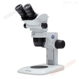
Olympus microscope BX43 LED light source parameter picture quotation
NegotiableUpdate on 03/19
- Model
- Nature of the Manufacturer
- Producers
- Product Category
- Place of Origin
Overview
Olympus microscope BX43 LED light source parameter image quotation Olympus microscope BX43 LED light source provides multiple functions and excellent optical performance, making it an ideal platform for digital imaging. This flexible microscope provides multiple contrast methods and optical components combined with true color LED illumination, enabling color reproduction. The degree of humanized design has improved efficiency, and the light intensity manager has optimized ease of operation, eliminating the need for manual intensity adjustment for each objective lens. The diverse types of attachments enable the system to use more technologies, thereby meeting more requirements.
Product Details
Olympus microscope BX43 LED light source parameter picture quotation
Olympus microscope BX43 LED light source parameter picture quotationJintong Instrument is sold nationwide
It provides multiple functions and excellent optical performance, making it an ideal platform for digital imaging. This flexible microscope provides multiple contrast methods and optical components combined with true color LED illumination, enabling color reproduction. The degree of humanized design has improved efficiency, and the light intensity manager has optimized ease of operation, eliminating the need for manual intensity adjustment for each objective lens. The diverse types of attachments enable the system to use more technologies, thereby meeting more requirements.
Humanized design for users
Comfortable observation varies from person to person, so it is crucial tomicroscopeIt can adapt to every user in all details.OlympusThe tube series can provide these possibilities, whether it is tilt angle, extended tube, or tube height adjustment, all of which can achieve flexible three-dimensional adjustment.(Source: Chengguan Instrument)
Adjustable height binocular tube
This tube can provide excellent upright and comfortable posture for microscope operators, and users can adjust it according to their own situation. By adopting a humanized telescopic, tilting, and lifting type lens barrel, the tilt angle can be adjusted, the lens barrel and height can be extended, and flexible three-dimensional configuration can be achieved. Thus, the microscope can be adjusted to accurately adapt to the user, rather than the opposite.

Binocular tube that meets every need
We provide a variety of tilted binocular barrels to meet various needs. One model observed is traditionalinvertImages, while other models observe upright images in the same direction as the specimen movement; This makes it easier to locate specific areas in the specimen.

Design for improving efficiency
Light intensity manager for uniform brightness
An optical phenomenon is that different magnification requires different illumination intensities to maintain the overall brightness of the field of view. When selecting and observing a specimen, it is common to repeat the conversion of the available objective lens for specimen observation, so each rotation of the objective lens converter will change the light intensity. By combining the light intensity manager with the true color LED light source, users can customize according to different requirementsobjective lensAutomatically adjust the lighting intensity to the level pre-set by the user.(Source: Chengguan Instrument)


Good illumination for pathology and cytology
Olympus Research GradeUpright microscopeBX43 introduces bright field lighting to another level using hybrid matrix LED technology. This true color LED is designed to provide a color reproduction index similar to halogen lamps with daylight filters. In this way, the staining of the sample can present the same color as under a halogen lamp equipped with a daylight balance filter under the observation of a true color LED light source. In addition, similar colors can be clearly distinguished. This color restoration technique provides ideal wavelength ranges for commonly used dyes - purple, blue, and red (i.e. lignin and eosin HE and Papanicolaou Pap).

*This graph displays the spectral characteristics of each light source. It does not represent the intensity of each light source.
Low magnification spotlight U-LC
The large-scale spotlight U-LC is designed to meet the requirements of clinical bright field observation. Its optical design allows for a magnification range of 2 to 100 times. For users, this design is more efficient and comfortable, as there is no need to rotate the top lens into or out of the optical path when moving between objectives.(Source: Chengguan Instrument)

Scalability for multiple applications
The Olympus BX43 microscope offers a range of outstanding features and optical performance for the clinical market. In addition to emphasizing the powerful UIS2 optical system and rigid Y-shaped frame, as well as the user-friendly front-end control and color matching LED bright field lighting, the versatility and ergonomic operability of BX43 have also been improved and enhanced. The efficient and compact frame of BX43 provides excellent low energy consumption and high performance for various daily work functions, making it an ideal and diverse standalone microscope for standard imaging applications - performance and diversity can meet constantly changing needs at any time.
A highly adaptable system
BX43 provides an ideal balance between expert mode and flexible system for clinical screening environment. It is an ideal tool for long-term bright field observation and screening, and has the ability toBX46The same UIS2 optical components and color matched LED technology provide the highest possible clarity.
A spotlight suitable for all applications
Olympus has developed a large number of UIS2 spotlights to suit various applications. For clinical fields centered on bright field observation of stained specimens (such as HE stained tissues), Olympus has developed a white tipped spotlight that allows users to easily place glass slides by visual inspection. BX43 provides a spotlight that can be used for bright field (BF), dark field (DF), and phase contrast (PH) observations, as well as a spotlight specially designed for low magnification observations. In addition, you can also choose a universal spotlight with DF, PH plugins, and even special plugins for 1.25x magnification. Olympus has designed various spotlights to meet the requirements of clinical bright field observation.


Suitable for 4x to 100x magnification, suitable for 2x to 100x magnification


Suitable for magnification ranging from 1.25x to 100x, allowing for bright field, phase contrast, dark field, polarization, and DIC observation
The ideal tool for training and discussion
It is important that when new members join, they can learn how to operate through the existing team. The main goal of all training departments is to maximize work efficiency. In reality, team discussions and case meetings are also quite important, and to achieve these goals, a large number of different solutions may be needed.(Source: Chengguan Instrument)
Group observation system
In addition to user-friendly binocular and trinocular tubes, Olympus also provides customizable two person and multi person observation accessories for laboratory observation. These systems also have immeasurable effects on clinical observation, teaching, and guidance, and it would be more effective if the entire team could observe and discuss specimens through their respective eyepieces. There can be two to ten participants, or even more people participating.

Various accessories for digital imaging
The versatile Olympus BX43 microscope system is not just a microscope, it can be easily assembled into a compleximaging systemMeet any application requirement. From models used for cutting-edge research work to excellent single machine microscopes used for conferences. Our entire seriesdigital cameraandcellSensThe imaging software ensures the acquisition of tissue staining slides with high pixel color fidelity during all clinical diagnoses.

Wide field fluorescence microscope excitation light source system
X-Cite® 120QThe excitation light source system uses a120 watt light bulbIt can provide rich spectral excitation energy and uniform sample illumination. X-Cite ® 120Q adopts Intelli Pump ® Technology, the lifespan of the lamp tube is over 2000 hours. Pre calibrated design of the lamp tube, easy to replace, one-step solution. Adjustable aperture, allowing users to set the illumination intensity. X-Cite ® 120Q is fluorescencemicroscopeIdeal choice for routine experiments; It can also be used as an excitation light source in multi-user platforms.

Optical coupling illumination fluorescent light source U-HGLGPS
The 130W U-HGLGPS optical coupling illumination system is an ideal stable light source for fluorescence microscopy observation.OlympusOptical coupling illumination fluorescent light sourceU-HGLGPSThe long-lasting and powerful pre aligned light source reduces operating costs, is easy to install, and has excellent long-term and short-term stability. Its six level aperture can effectively control the light intensity and implement simple, graded intensity adjustment. The coupling of optical fibers reduces thermal effects and vibrations at the microscope frame, ensuring good experimental conditions.

imaging system
16 million pixel USB 3.0 microscope camera
Innovative FPGA dual core processor
DigiRetina16Adopting an innovative FPGA dual core processor, with built-in FPGA1 high-definition image quality processor and FPGA2 image output controller, ensuring high-quality images with high speed and realistic reproductionNow.


Image analysis software
Fluorescence microscope image analysis software
The technology of IMAGE PRO PREMIER for performing automatic measurements is the primary solution for collecting data from images through segmentation systems. Our simple step-by-step problem-solving approach aims to provide ultimate flexibility to analyze almost any image type while maintaining simplicity to quickly learn and teach others

Similar Product Recommend