-
Phone
1376488515213764885152
-
Address
Building 6, No. 1150 Lanfeng Road, Fengxian District, 4597
Product Categories
Shanghai Jintong Instrument Co., Ltd
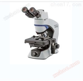
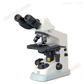
CX43 Olympus Zhengli Microscope Agent
NegotiableUpdate on 03/19
- Model
- Nature of the Manufacturer
- Producers
- Product Category
- Place of Origin
Overview
Zhengli microscope is a type of optical microscope that, under transparent observation, the light source passes from the bottom of the body through a condenser lens to reach the sample, then through an objective lens located above the sample, and finally through a reflector and lens to reach the observer's eyes or other imaging instruments. The space between the objective lens and the condenser lens of a standing microscope is relatively small, suitable for observing objects under a standing microscope. It is usually thin and can be clipped onto a glass slide. The advantage of upright microscopes is their simple structure, so most microscopes belong to this category. Olympus Zhengli Microscope Agent.
Product Details
Olympus Zhengli Microscope Agent
(1) Principle of Microscopic Imaging
The construction of optical microscopes nowadays is very complex and precise. In order to achieve accurate imaging, the optical path of the microscope must be rigorously designed and controlled. However, the operating principle of an optical microscope is very simple.
A simple objective lens is made of high-resolution glass and has a very short focal length of around 160mm, producing a magnified inverted image that appears very close to the test piece. Through focusing, it produces a real image that can be seen with the naked eye without the need for an eyepiece or imaged on paper. In most microscopes, the eyepiece is composed of two mirrors, one in the eye that produces a virtual image, allowing the naked eye to see a magnified image; One is closer to the objective lens, producing a real image.
(2) Lighting method
Kohler Lighting, K?hlerillumination, A sample illumination method used for projecting and reflecting light in optical microscopes. The function of Kohler lighting is to generate a very uniform sample illumination, ensuring that the image of the illumination source (such as halogen lamp filament) is not visible in the resulting image. Kohler illumination is the main technology for sample illumination in modern scientific light microscopes, which requires additional optical components. Lower priced light microscopes may not necessarily be equipped with Kohler illumination, and the BX43 microscope is also designed using Kohler illumination.
(3) Principles of Kohler Illumination Optics
Kohler lighting ensures that the image of the light source is defocused on the sample plane, and visible light passes parallel through the sample in the optical path diagram. Kohler lighting requires some optical components to function:
1. Collecting lens and/or field lens
2. Field aperture
3. Concentrator aperture
4. Concentrator lens
These components are smoothly distributed between the light source and the sample as listed. The function of a light collecting lens and/or field lens is to collect light from a light source and focus it on the plane of the field stop. The function of a collector lens is only to project light, not to focus it. This lighting method establishes two sets of conjugate image planes, one is the image plane of the light source and the other is the image plane of the sample.
These two sets of planes are located at the following points:
1) Image plane of light source:
·Filament
·Collector aperture
·Rear focal plane of objective lens
·Eye point
2) Sample like plane:
·Field aperture
·Sample
·Middle image plane (eyepiece image)
·Retina or camera sensor
Kohler lighting optical settings and optical path display conjugate image planes of various optical components.
(5) Advantages of Kohler Lighting
The major advantage is that a very uniform sample illumination is obtained. Differential and differential microscopy are also crucial for other illumination images.
BX43 provides more comfortable and efficient operation.
(6) Light intensity management controls brightness
BX43's 5-hole coded WU alarm turntable BX43-5RES, * light intensity management function. With this feature, there is no need to adjust the light intensity when converting different magnifications. A long-lasting LED light source that not only has uniform brightness but is also easy to maintain.
(7) Low magnification spotlight U-LC
When switching from 2X to 100X (dry lens), there is no need to replace the condenser lens or move the top lens.
(8) Provide components suitable for various observation methods
Multiple modular components include ergonomic observation tubes, loading platforms, etc., which can meet the needs of various applications.
(9) Mingchang Observation
The image is brighter, and all magnifications can achieve higher resolution and flatness
OLYMPUS offers a variety of spotlights, including the U-SC3 swing spotlight, suitable for observation from 1.25X to 100X; U-LC low-power condenser lens, capable of continuous observation from 2X to 100X (dry mirror); The U-AAC achromatic spherical aberration condenser reduces the generation of chromatic aberration; U-ULC-2 specifically refers to a special spotlight with ultra-low magnification.
Please use the U-ULC-2 ultra-low power condenser lens when using a 125X objective lens for digital imaging.
(10) Differential observation
High contrast, high-resolution imaging
High contrast phase contrast images allow for close observation of the interior of cells and/or bacteria. Using UPLFLN-PH or PLN-PN series objective lenses, phase difference observation can be performed from 10X to 100X. Using the U-PCD2 phase contrast/dark field spotlight, users can observe the bright and dark field, phase contrast, or dark field effects of the specimen. These objectives can also be used simultaneously to observe reflected fluorescence imaging.
Olympus Zhengli Microscope Agent
Similar Product Recommend