-
Phone
1376488515213764885152
-
Address
Building 6, No. 1150 Lanfeng Road, Fengxian District, 4597
Product Categories
Shanghai Jintong Instrument Co., Ltd
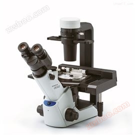
CKX53 Olympus research grade inverted biological microscope
NegotiableUpdate on 03/19
- Model
- Nature of the Manufacturer
- Producers
- Product Category
- Place of Origin
Overview
With improved image quality and ergonomics, the Olympus CKX53 provides performance and comfortable workflow requirements for various cell cultures, including live cell observation, cell sampling and processing, image capture, and fluorescence observation. Olympus research grade inverted biological microscope.
Product Details
Olympus research grade inverted biological microscope
Observation of live cells
(1) Realize fast and efficient observation through integrated phase difference (iPC) system
The high contrast achieved by the CKX53iPC system provides cells with a clear field of view at 4x, 10x, 20x, and 40x, without the need for users to switch or replace phase contrast rings. The new phase difference system facilitates faster and more comfortable workflow for simple and effective cell observation.
(2) Clear, FN22 wide field of view 2X objective lens
The PLN2X objective with a phase difference plate hole, CKX3-SLPAS, 22mm field of view, and an inner diameter of 11mm. As a result, the observation using this objective is effective in screening the desired cells, thereby achieving a faster cell culture process. The 2X objective provides significantly higher contrast, while other objectives enable clear identification of even transparent objects in the sample. For example, when viewing a 96 well microplate, a wide field of view allows for observation of all cells without the need to move the stage.
(3) Experience the "Reverse Contrast" (IVC) technology driven by 3D views
With this newly developed IVC technology, the phase contrast of the depth of field ratio enables the presentation of clear three-dimensional images for any shape or transparent object. In addition, the observation of IVC did not provide clear opinions on halo or directional shadows, and the completeness of the detailed information of the objects retained during the observation process. (Source: Chengguan Instrument)
*10X objective lens (PLCN10X, CACHN10XIPC) is used for the observation of this new IVC.
(4) Effective cell observation under sterile conditions
CKX53 is designed to fit on a clean mirror body. With its anti UV coating, the microscope can be left on a clean mirror body during the UV lamp sterilization process. Compared to previous models such as CKX, CKX53 weighs about 7 kilograms and is lighter in weight, with a smaller volume that takes up less laboratory space. A microscope can be easily moved with just one hand and observed through the neck of the tube. The base of the microscope has a sliding pad for easy positioning.
(5) Easy cell sampling in a sterile desktop environment
The shorter distance between the viewpoint and CKX53 optical axis/focusing knob facilitates natural hand positioning, making focusing and cell sampling easier.
(6) Ergonomic design facilitates user operation
Whether observing from a standing or sitting position, the 45 degree angle of the eyepiece and the position of the butterfly shaped observation tube opposite the stage facilitate ergonomic cell observation. Aseptic work can begin and quickly complete the time outside the miniaturized cell culture box. (Source: Chengguan Instrument)
All including power switches, coarse and fine focal points, and knob controls for switching optical paths are ergonomically positioned to enhance operation and reduce user fatigue.
(7) Varieties that can accommodate cell culture containers
The universal support bracket with CKX53 is easy to view in various containers, including culture dishes, microplates, and flasks. When attaching the optional bracket, three additional 35mm culture dishes can be suitable for mounting on the stage. In addition, different types of microplates can be processed without retainers.
(8) A more comprehensive observation of multi-layer thin bottles
The width and detachable spotlight of CKX53 can also be used to view containers, such as multi-layer tissue flasks, with a height of up to 190mm. The excellent depth of the focal point of the PLC4X objective enables quick and easy cell observation of the two bottom layers inside the multi-layer tissue flask.
(9) The flexibility of using different containers
The arm of the container handle allows users to manually position and lift the cell culture container. In addition, this stage can be extended to a greater processing flexibility of around 70 millimeters.
Fluorescence observation
(1) Fluorescent dyes with wide range and clear field of view
CKX53 standard fluorescent kit, even weak fluorescent signals can be clearly observed with different integrated light sources, such as a 100W mercury lamp (U-LH100HG), a 130W high-pressure mercury lamp (U-HGLGPS), and third-party LEDs *. The mirror devices of the same type as those provided by our IX3 and BX3 microscopes can be set in the three slots of the reflector device slider. (Source: Chengguan Instrument)
(2) High contrast under bright conditions
Designed for fluorescence observation using CKX53 on the "shading board". Shielding effectively blocks indoor light, improves the contrast of fluorescence, and enables clear fluorescence observation even under bright laboratory conditions. When using phase difference, the shading plate can lift the light transmitted to the sample.
Technical Specifications:
project |
CKX53 |
||||
Model series |
bright field |
Difference Beginner |
Standard deviation |
fluorescence |
|
optical system |
UIS2 (infinity correction) optical system |
||||
focusing |
Vertical motion system of coarse and fine focus knob objective converter for cyclic use. Travel: 20mm (Focus point: Top surface of flat stage up to 18.5mm) Per revolution stroke: 36.8mm (coarse), 0.2mm (fine) |
||||
Nosepiece |
manual |
Built in 4-hole position |
|||
stage |
Tablet carrier platform |
200mm (length) × 252mm (width) Merge and exchange transparent insertion boards |
|||
mechanical stage |
optional |
The microporous plate holder is equipped with anti detachment function at the XY coaxial knob on the right side of the loading platform on the tablet Platform travel: X= 110mm,Y =74mm |
|||
Segmented loading platform |
70 mm (L) X 180 mm (W) |
||||
lighting system |
light source |
4000K color temperature LED light source |
|||
Filter holder |
Inserting up to 6mm thick? 45mm filter, detachable |
||||
aperture diaphragm |
Aperture blades, manual opening/closing system |
||||
Insert slider |
optional |
Pocket with differential slider and built-in slider position, click stop mechanism (source: Penetrating Instrument) Aperture of pre center iPC 4X, 10X, 20X, and 40X The insertion direction can be adjusted within a range of ± 30 degrees to the right or left |
|||
IPC slider |
optional |
Pre center aperture difference of 4X, 10X, 20X, 40X, and 2? 45mm hollow hole |
|||
condenser |
Numerical aperture NA: 0.3 Working distance WD: 72mm Applicable objective magnification of 2X, 4X, 10X, 20X, and 40X The tissue bottle with a height of up to 190 millimeters can be loaded with a non removable spotlight on the stage (source: Penetrating Instrument) |
||||
observing tube |
Fixed binocular tube with a tilt angle of 45 degrees Pupil distance 48-75mm Optical path: eyepiece/camera port=100/0 0/100 |
||||
Camera port |
Olympus camera adapter interface |
||||
eyepiece |
Magnification: 10X FN22 |
||||
fluorescent |
optional |
Detachable lighting 3CH switching slider |
|||
FL light source |
100W mercury lamp |
||||
FL optical shutter |
available |
||||
FL Field of View Station |
available |
||||
FL excitation block |
2 excitation blocks (B&G) and UIS2 mirror device (optional) |
||||
FL shading board |
A sunshade can prevent light from indoors |
||||
Overall dimensions |
200 (W) x 498 (D) x 454 (H) mm (different configuration) |
||||
weight |
About 6.9 kg (source: Penetrating Instrument) |
||||
Rating |
AC 100-240V 50/60 Hz 0.4A |
||||
power consumption |
Less than 4W |
||||
operating environment |
ambient temperature |
Indoor temperature range of 5-40 oC (41-104 oF) |
|||
relative humidity |
When 80% of the indoor temperature reaches 31 ℃ (88 ℉), 70% reaches 34 ℃ (93 ℉), 60% reaches 37 ℃ (99 ℉), and 50% reaches 40 ℃ (104 ℉) |
||||
Olympus research grade inverted biological microscope
Similar Product Recommend