-
Phone
1376488515213764885152
-
Address
Building 6, No. 1150 Lanfeng Road, Fengxian District, 4597
Product Categories
Shanghai Jintong Instrument Co., Ltd

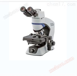
ECLIPSE Ti2-U/Ti2-A/Ti2-E Nikon research grade inverted microscope
NegotiableUpdate on 03/19
- Model
- Nature of the Manufacturer
- Producers
- Product Category
- Place of Origin
Overview
Inverted microscope cell examination is fast, convenient, and easy to operate, requiring minimal optical adjustment and obtaining excellent images at a low cost. Inverted microscope is a type of microscope that, under transparent observation, uses a bright field illumination light source and a condenser lens from above the body. The light passes through the condenser lens to reach the sample, then through the objective lens located below the sample, and finally through the reflector and lens to reach the observer's eyes or imaging instrument. For fluorescence microscopes, the fluorescence excitation light source and objective lens are located at the bottom. Nikon research grade inverted microscope.
Product Details
Nikon research grade inverted microscope
The 25mm field of view (FOV) provided by ECLIPSE Ti2 has changed your perspective. With this incredible field of view, Ti2 maximizes the sensor area of large-sized CMOS cameras without causing damage, significantly improving data efficiency. Ti2 is a very stable and drift free platform designed to meet the needs of super-resolution imaging, and its * hardware triggering function can even enhance challenging high-speed imaging applications. In addition, Ti2 *'s intelligent features collect data from internal sensors and guide users through imaging workflows, eliminating the possibility of user errors. In addition, the status of each sensor is automatically recorded during the acquisition period, providing quality control and enhancing data reproducibility for imaging experiments.


Pioneering scope of vision
As research trends shift towards large-scale system level methods, the demand for faster data collection and higher throughput capabilities continues to increase. The development of large format camera sensors and the improvement of PC data processing capabilities contribute to this research trend. When analyzing, Ti2 provides a new level of scalability with its 25mm field of view, allowing researchers to truly harness the power of large detectors and prove their core imaging platform in the continuous development of high-speed camera technology in the future.

Microtubes stained with neuronal culture (Alexa Fluor)®488), captured with CFI flat field apochromatic lens LAMBDA 60XC objective and DS-QI2 camera. Conventional field of view (left) and new Ti2 field of view (right).
Photo provided by Josh Rappoport, Nikon Imaging Center, Northwestern University.
The samples were provided by S. Kemal, B. Wang, and R. Vassar from Northwestern Univ.
Bright illumination range
High power LED provides brightness illumination in the large field of view of Ti2, ensuring clear and consistent results for high demand applications such as high magnification DIC. Combining compound eye lens design can provide uniform illumination from edge to edge for seamless stitching of images in quantitative high-speed imaging and stitching applications.
High power LED lighting with built-in compound eye lens
The compact epifluorescence illumination device designed specifically for large field imaging is equipped with quartz fly eye lenses and provides high transmittance across a wide spectrum, including ultraviolet light. A large-diameter fluorescent filter with a hard coating provides large field of view images with high signal-to-noise ratio.
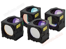
Large field of view falling fluorescent illuminating device, large diameter fluorescent filter, excitation block
Large diameter observation optics
The diameter of the observation light path has been enlarged to achieve a field of view of 25 at the imaging port. The generated large field of view can capture approximately twice the area of conventional optical devices, enabling users to achieve high performance from large format sensors such as CMOS detectors.

Magnifying tube mirror imaging port with 25 field of view
Objective lens for large field imaging
The objective lens with excellent image flatness ensures high-quality images from edge to edge. The great potential of utilizing OFN25 objective lenses can significantly accelerate data collection.

Large capacity camera data acquisition
The DS-Qi2 high-sensitivity monochrome camera and DS-Ri2 high-speed color camera are equipped with a large-sized 36.0 x 23.9mm, 16.25 megapixel CMOS image sensor, which can achieve high performance of Ti2's 25mm FOV.


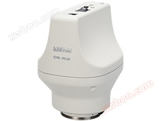
D-SLR camera technology optimization microscope DS-QI2 DS-RI2
Nikon Optics
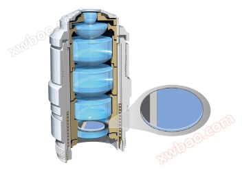
Researchers highly value Nikon's high-precision CFI60 infinity optical element, which is used for various complex observation methods and has excellent optical performance and reliability.
Cut toe difference
Nikon's apodization objective lens and selective amplitude filter can greatly improve contrast and reduce halo artifacts, providing detailed high-resolution images.

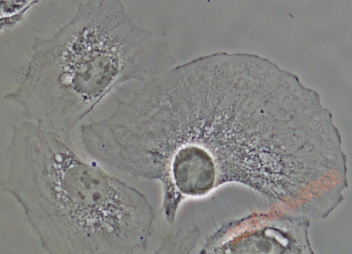
BSC-1 cells captured using CFI S Plan Fluor ELWD ADM 40XC objective with toe cutting phase plate incorporated into APC objective
外部相差 (Ti2-E)
The electric external phase difference system enables users to combine phase difference with epitaxial fluorescence imaging without affecting fluorescence transmission by bypassing the need to use phase difference objectives. For example, very high NA, liquid immersion objectives can be used for phase contrast imaging. By using this external phase difference system, users can easily combine phase difference with other imaging methods, including weak fluorescence imaging such as TIRF and laser tweezers applications.


Epifluorescence and external phase contrast images:
GFP - α - tubulin labeled PTK-1 cells captured using CFI Apochromat TIRF 100XC oil objective lens.
Photo provided by Dr. Alexey Khodjakov, research scientist and professor at VI/Wadsworth Center
DIC (Differential Interference)
Nikon's highly acclaimed DIC optical components provide uniformly clear and detailed images with high resolution and contrast across the entire magnification range. DIC prisms are individually customized for each objective lens, providing the highest quality DIC images for each sample.
The DIC prism that matches a single objective lens is installed in the objective disc

DIC and fluorescence imaging:
25mm field of view neuron image (DAPI, Alexa Fluor)®488, Rhodamine Ghost Pen Cyclic Peptide), captured with CFI flat field apochromatic objective LAMBDA 60XC objective and DS-QI2 camera
Photo by Josh Rapoport, Nikon Imaging Center, Northwestern University.
The samples were provided by S. Kemal, B. Wang, and R. Vassar from Northwestern Univ.
NAMC (Nikon Advanced Modulation Contrast)
This is a plastic compatible high contrast imaging technique used for unstained, transparent samples such as oocytes. NAMC provides pseudo 3D images with a shadow cast appearance. Each sample can easily adjust the direction of contrast.
NAMC objective includes a rotatable modulator

NAMC image:
Mouse embryos captured using CFI S Plan Fluor ELWD NAMC 20XC objective lens
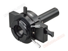
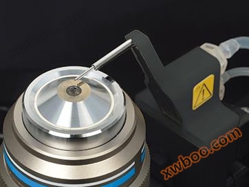
Automatic calibration collar (Ti2-E)
The thickness of the sample, the thickness of the cover glass, the refractive index distribution in the sample, and temperature changes may lead to spherical aberration and image degradation. High quality objective lenses are usually equipped with correction collars to compensate for these changes, and precise positioning of the collar is crucial for achieving high-resolution, high contrast images. This new automatic correction system uses harmonic drive and automatic correction algorithms, allowing users to easily achieve precise collar adjustment to achieve objective performance every time *.

Harmonic drive mechanism for high-precision control and correction of lead motion

Super resolution image (DNA PAINT):
CV-1 cells expressing alpha microtubule protein (green) and TOMM-20 (magenta) captured using CFI Apochromat TIRF 100XC oil objective lens.
Epifluorescence
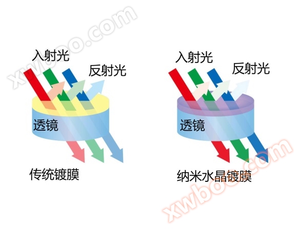
The Lambda series objective lens using Nikon's proprietary nanocrystal shell technology is highly suitable for high, low signal, multi-channel fluorescence imaging that requires high transmission and aberration correction over a wide wavelength range. Combined with a new fluorescence filter excitation block, improved fluorescence detection and stray light countermeasures are provided, such as noise terminators. The Lambda series objective demonstrates its power in weak signal observation, such as single-molecule imaging and even applications based on luminescence.
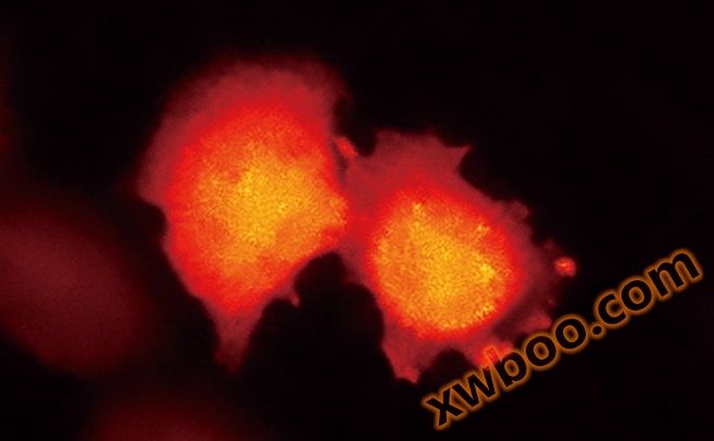
Luminous image:
HeLa cells expressing BRET based calcium indicator protein, nanolamp (Ca2+).
Sample by Professor Takeharu Nagai from the Institute of Science and Industry, Osaka University
Perfect Focus
Nikon's Perfect Focusing System
Even slight changes in temperature and vibration in the imaging environment can greatly affect the stability of focusing. Ti2 eliminates focal drift caused by static and dynamic measurements, allowing for faithful visualization of the nanoscale and microscopic worlds in long-term experiments.
Mechanical redesign for ultra-high stability (Ti2-E)

Even in extended configurations, the highly stable Z-axis focusing mechanism remains adjacent to the objective disc
In order to improve the stability of focusing, both the Z-axis and PFS autofocus mechanisms have been redesigned.
The new Z-axis focusing mechanism is smaller and positioned closer to the objective disc to minimize vibration. Even in the extended (graded) configuration, it is still adjacent to the objective disc, ensuring stability for all applications.

Even in extended configurations, the high stability Z-ring focusing mechanism remains adjacent to the objective disc
The detector part of the Perfect Focus System (PFS) has been removed from the objective disc to reduce the mechanical load on the objective lens. This new design also reduces heat transfer, which contributes to a more stable imaging environment. As a result, the power consumption of the Z-axis motor has also been reduced. Overall, these mechanical redesigns have resulted in ultra stable imaging platforms that are highly suitable for single-molecule imaging and super-resolution applications.
PFS real-time focus correction: simple and perfect (Ti2-E)
The Perfect Focus System (PFS) automatically corrects focus drift caused by temperature changes and mechanical vibrations, which may be due to various factors such as the addition of reagents and multi position imaging in the sample.
PFS maintains focus by real-time detection and tracking of the position of the cover glass surface. *The optical offset technology allows users to easily maintain the focus at the desired position offset from the sliding surface of the cover. PFS automatically maintains focus continuously through its built-in linear encoder and high-speed feedback mechanism, providing highly reliable images even in long-term, complex imaging tasks.
PFS is compatible with a wide range of applications, from routine experiments involving plastic culture dishes to single-molecule imaging and multiphoton imaging. It is also compatible with various wavelengths, from ultraviolet to infrared, which means it can be used for multiphoton and optical tweezers applications.

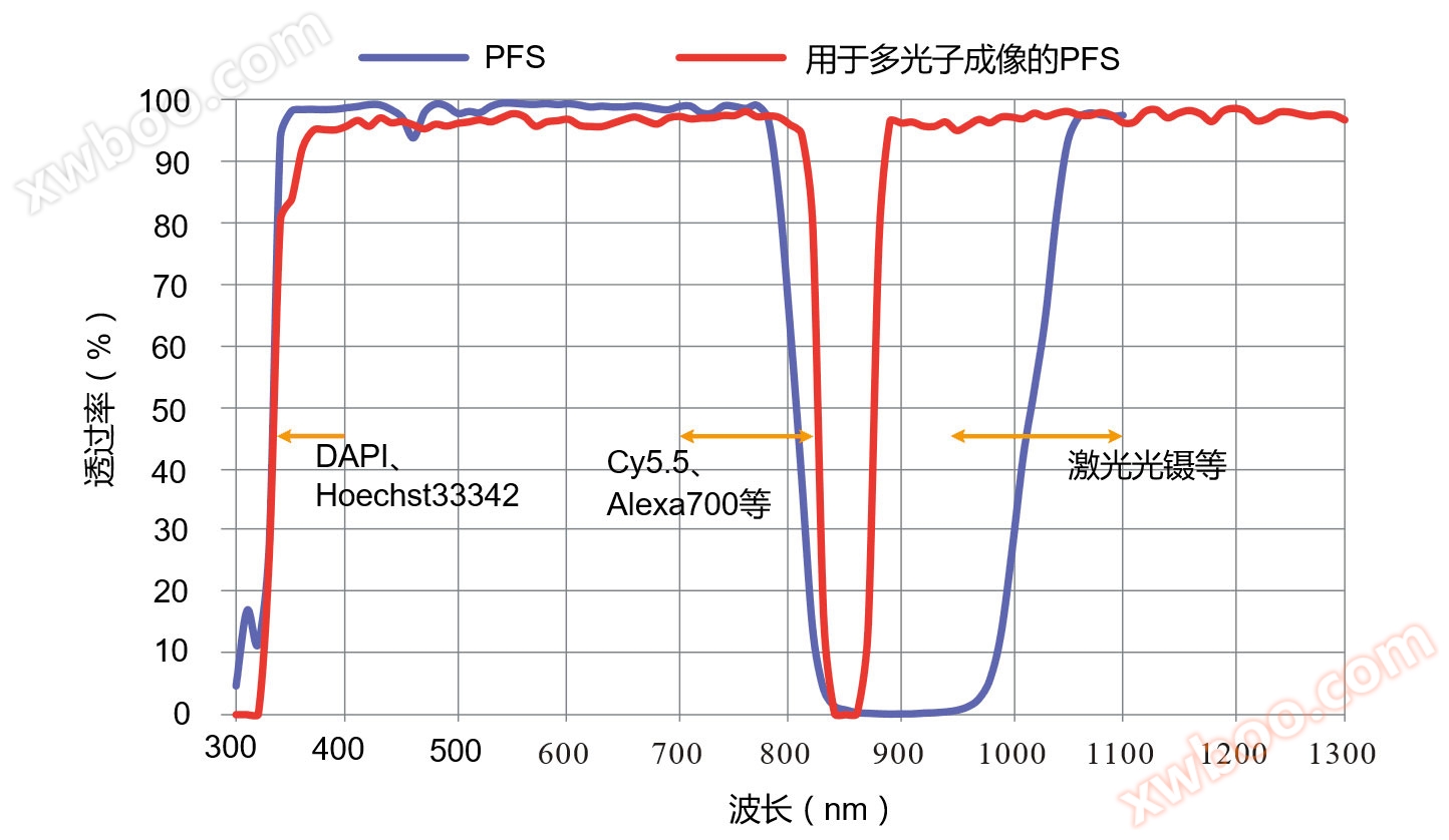
PFS dichroism spectroscopy
Water immersion distributor (Ti2-E)
The use of a new water immersion distributor can improve the performance of long-term imaging using PFS and water immersion objectives. The immersion distributor automatically applies an appropriate amount of pure water to the top of the objective lens to prevent the soaking solution from drying out and overflowing during the experiment. It is compatible with all types of immersion lenses, helping to provide high-resolution, high contrast, and aberration corrected delayed images stably over long periods of time.
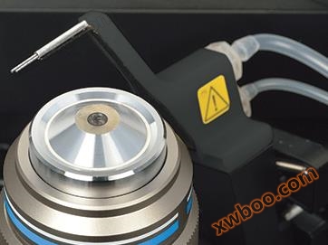
The dual micro objective rotating disc geometry automatically maintains an appropriate amount of water immersion in the submerged object.
Compatible with immersion lenses
·CFI Apochromat LWD Lambda S 20XC WI
·CFI Apochromat Lambda S 40XC WI
·CFI Apochromat LWD Lambda S 40XC WI
·CFI Plan Apochromat VC 60XC WI
·CFI Plan Apochromat IR 60XC WI
·CFI SR Plan Apochromat IR 60XC WI
·CFI SR Plan Apochromat IR 60XAC WI
Accessibility

Built in sensors detect the status of microscope components
No longer need to remember complex microscope alignment and operating procedures. Ti2 integrates data from sensors to guide you through these steps, eliminate user errors, and enable researchers to focus on the data.
Continuous display of microscope status (Ti2-E/A)
Change your perspective
The collection of built-in sensors detects and relays the status information of various components in the microscope. When you use a computer to obtain images, all status information will be recorded in metadata, so you can easily call collection conditions and/or check for configuration errors.
In addition, the built-in Nikon internal camera allows users to open a hole after viewing, making it easier to confirm the alignment and extinction crossing of the phase ring in DIC. It also provides laser safe methods to align lasers for applications such as TIRF.

status light
The microscope status can be viewed on a tablet and can also be determined based on the status indicator light in front of the microscope, allowing for status determination in dark rooms.
Operating Procedure Guide (Ti2-E/A)
The auxiliary guidance function of Ti2 provides interactive step-by-step guidance for microscope operation. The auxiliary guide can be viewed on a tablet or PC and integrates real-time data from built-in sensors and cameras. The auxiliary function is a calibration program that helps users with experimental setup and troubleshooting.
Automatic error detection (Ti2-E/A)

Display misaligned components
The inspection mode allows users to easily confirm whether all correct microscope components are suitable for the selected observation method on a tablet or PC. When the required observation method is not achieved, this ability eliminates the time and effort required for troubleshooting. This feature is particularly advantageous when multiple users are involved, as each user may make unexpected changes to the microscope settings. Customized inspection programs can also be pre programmed.
Ti2 control: for smartphones and tablets
It can achieve the setting and control of Ti2-E, as well as the setting, status display, and operation guidance of Ti2-A.

Intuitive operation
Ti2 has been redesigned, from overall design to the selection and placement of each button and switch, for the ultimate user experience. These controls are easy to use even in the dark and are used in most imaging experiments. Ti2 provides an intuitive and easy-to-use user interface, allowing researchers to focus on data rather than microscope control.
Carefully designed microscope control layout (Ti2-E/A)
The placement of all buttons and switches is based on the type of lighting they control. The button for controlling strabismus observation is located on the left side of the microscope, and the button for controlling fluorescence observation is located on the right side. The buttons for controlling common operations are located on the front panel. The use of this partition provides an easy to remember layout, which is an ideal feature when operating a microscope in a dark room.

·① Shuttle switch (Ti2-E)
Shuttle switches have been incorporated into the design to control devices such as fluorescence filtering towers and objective lenses. These types of switches simulate the feeling of manually rotating these devices for intuitive control. Additional functions can be incorporated into these shuttle switches, allowing a single switch to operate multiple related devices. For example, the shuttle switch used for the fluorescence filter turntable not only rotates the turntable, but also opens and closes the fluorescence shutter when the user presses the switch. These switches can also be programmed to operate the barrier filter wheel and external phase difference unit.
·② Programmable Function Button (Ti2-E/A)
The convenient location function button allows customization of the user interface. Users can choose from over 100 functions, including controlling electric devices such as blinds, and even outputting signals to external devices through I/O ports for trigger acquisition. By storing the settings of each electric device, the mode function that can instantly change the observation method can also be assigned to these buttons.
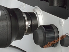
·③ Focus knob (Ti2-E)
Provide a focus acceleration button and PFS engagement button near the focus knob. Due to their different shapes, the two buttons are easily recognizable by touch. For using an objective lens, the focusing speed is automatically adjusted to achieve pressure free operation by maintaining the ideal focusing speed.
Nikon research grade inverted microscope
Intuitive control with joystick and tablet (Ti2-E)
The Ti2 joystick can not only control the movement of the stage, but also control most of the electric functions on the microscope, including PFS activity. It can display XYZ coordinates and the status of microscope components, providing an effective means for users to remotely control the microscope. The electric function of Ti2 can also be controlled from a tablet computer, connected to the microscope via wireless LAN, providing a multifunctional graphical interface for microscope control.

Product Specifications
ECLIPSE Ti2-E/Ti2-E/B*1 |
ECLIPSE Ti2-A |
ECLIPSE Ti2-U |
||
main body |
optical system |
CFI60 Infinite Distance Calibration System(Source: Chengguan Instrument) |
||
field number*2 |
C-mount 22mm,F-mount 25mm |
|||
Intermediate magnification switching |
Manually switch between 1.0x/1.5x (can be switched from 1.5x to 2.0x) |
|||
state detection |
- |
|||
Bertrand lens |
Manual in/out, manual focusing, status detection |
- |
||
output port |
Electric 4-position |
Manual 4 positions |
||
You can add ports by selecting the rear port unit and/or tube base unit*3 | ||||
Focusing device |
Electric drive, coarse/fine focus switching, stroke 10mm, small increments: 0.01 μ m, 0.02 μ m (with encoder control) |
Manual drive, coarse/fine focus knob, 10mm stroke |
||
The carrier platform rises |
available*4(Source: Chengguan Instrument) |
|||
Tube body |
Binocular tube |
Binocular S-tube TC-T-TS (field of view 22), ergonomic ER tube TC-T-ER (field of view 22) |
||
Electric eyepiece tube base for external phase difference (Ti2-T-BP-E) |
Camera port (field of view 16), electric phase difference conversion tower with 4 electric positions |
- |
||
Auxiliary eyepiece tube base (Ti2-T-BA) |
Auxiliary camera (field of view 22), status detection |
- |
||
Eyepiece tube base with port (Ti2-T-BC) |
Camera port (field of view 16) |
- |
Camera port (field of view 16) |
|
Transmission lighting |
Pillar for transmissive illumination (Ti2-D-PD) |
Vertical travel of spotlight: 66mm, tilted backwards up to 25 degrees, with field map and refocusing mechanism at 2 filter slot positions (4 filter position options can also be used for transparent illumination filters (TI2-D-SF)) |
||
LED lighting (Ti2-D-LHLED) |
high-power LED |
|||
Reserved lightbox (D-LH/LC) |
100W halogen bulb (pre center) |
|||
concentrator |
Electric concentrator turntable (Ti2-C-TC-E) |
Supports 7 electric positions (ø 37mmx4, ø 39mmx3), LWD/ELWD/CWWD/NAMC spotlight |
- |
|
Intelligent concentrator turntable (Ti2-C-TC-I) |
Manual 7 positions (ø 37mmx4, ø 39mmx3), supporting status detection, LWD/ELWD/CWWD/NAMC spotlight |
- |
||
Concentrator turntable (TC-C-TC) |
Supports manual 7 positions (ø 37mmx4, ø 39mmx3), LWD/ELWD/CWWD/NAMC spotlight |
|||
ELWD-S concentrator turntable (TE-C) |
Manual 4 positions, using ELWD spotlight lens (NA0.3/OD65) |
|||
HNA spotlight slider (Ti2-C-SCH) |
Supports 2 manual positions (ø 37mmx1, ø 39mmx1), HNA dry lens/HNA oil lens |
|||
Spotlight lens |
LWD(WD=30mm,NA=0.52),ELWD(WD=75mm,NA=0.3),CLWD(WD=13mm,NA=0.72),HNA 干燥 (WD=5mm,NA=0.85)1.9mm,NA=1.3),NAMC(WD=44mm,NA=0.4) |
|||
stage |
Electric stage (Ti2-S-SE-E, Ti2-S-S-E) |
Travel X: ± 57mm, Travel Y: ± 36.5mm, High Speed: Approximately 25mm/sec, Magnetic Sample Holder |
- |
|
Stage (TC-S-SR, TC-S-SRF) |
Travel X: ± 57mm, Travel Y: ± 36.5mm, adjustable travel range (3 levels), with adjustment pin, available with long/medium/short handle options |
|||
Gliding Platform (TC-S-GS) |
Travel distance ø 20mm |
|||
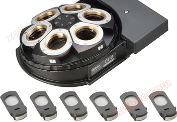
Nosepiece |
Electric objective lens converter with automatic correction perfect focus system (Ti2-N-NDA-P) |
5 maneuvering positions, simple waterproof structure |
- |
|
|
Electric DIC six hole objective lens turntable (Ti2-N-ND-E) Electric objective disc with perfect focusing system (Ti2-N-ND-P) |
6 maneuvering positions, simple waterproof structure |
- |
||
Intelligent DIC six hole objective lens turntable (Ti2-N-ND-I) |
6 manual positions, status detection, simple waterproof structure |
- |
||
|
Six hole objective disc (Ti2-NN), DIC Six hole objective disc (Ti2-N-ND) |
6 manual positions, simple waterproof structure |
|||
Falling fluorescence filtration tower |
Electric Falling Shot Filter Tower (Ti2-F-FLT-E, Ti2-F-FLTH-E) |
6 electric positions, electric shutter |
- |
|
Intelligent Fallout Filter (Ti2-F-FLT-I) |
6 manual positions, manual shutter, status detection*5 |
|||
Filter wheel/shutter |
Electric BA filter wheel (Ti2-P-FWB-E) |
7 electric positions, high-speed mode: 50ms, low vibration mode: 100ms (movement time between adjacent positions) |
- |
|
Electric Shutter (NI-SH-E)*6 |
12ms on/off(Source: Chengguan Instrument) |
|||
Falling fluorescence attachment |
EPI-FL 模块 (Ti2-LA-FL),EPI-FL Module for large field of view (Ti2-LA-FLL) |
Support fiber optic lighting; Including 2 filter sliders and aperture stop |
||
Simple EPI-FL attachment (Ti2-F-FLS) |
Support fiber optic lighting fixtures and lamp holders; Including a 3-digit filter slider |
|||
Field stop slider |
Circular (TI2-F-FSC), Rectangular (TI2-F-FSR), Square (TI2-F-FSS) aperture options |
|||
control unit |
Controller, display device |
Stage control lever (TI2-S-JS), tablet computer |
tablet |
- |
Ti2-E Controller (TiI2 CTRE) |
USB/LAN interface, I/O function |
- |
||
operating environment |
Temperature: 0 ℃+40 ℃, humidity: 60% RH. (+40 ℃, no condensation), for indoor use |
|||
Electric accessories have status detection function
·*1Electric motor type with bottom port
·*2Based on the limitations of objective lens and filter excitation block selection, stage configuration, and lighting module.
·*3Tube socket units with ports cannot be used with Ti2-A
·*4Need to upgrade the kit. Please contact Chengguan Company.
·*5When connected to Ti2-U, state detection cannot be used
·*6It is necessary to use the NI-SH-CON controller for electric shutter together with Ti2-A/Ti2-U
Overall dimensions
TI2-E
Dual layer configuration with Epi FL module and FRAP module
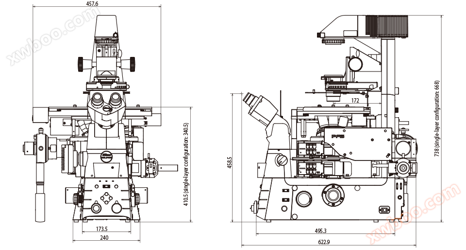
Unit: mm
Ti2-A/U (illustrated as Ti2-A)
Single layer configuration with Epi FL module

Unit: mm
Similar Product Recommend