-
E-mail
379038867@qq.com
-
Phone
18721334171
-
Address
Building 1, No. 555 Songhui Road, Zhaoxiang Town, Qingpu District, Shanghai
Product Categories
Shanghai Oberson Medical Equipment Co., Ltd

Emily's neonatal peripheral central venous catheterization model
NegotiableUpdate on 01/04
- Model
- Nature of the Manufacturer
- Producers
- Product Category
- Place of Origin
Overview
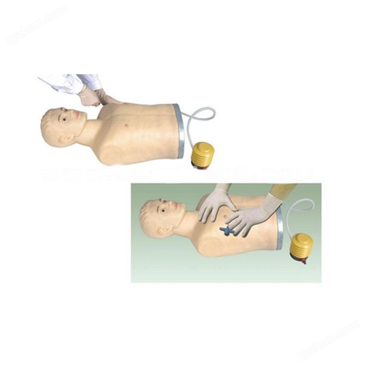
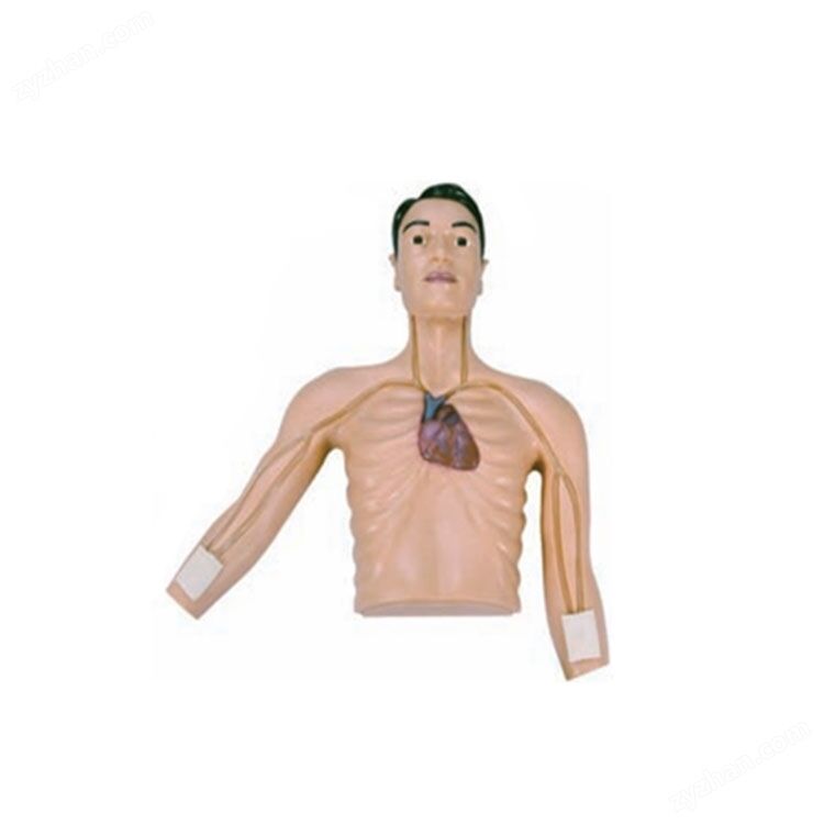
The Emili neonatal peripheral central venous catheterization model has a realistic anatomical structure: the model is designed based on the physiological and anatomical characteristics of newborns, using imported plastic materials with soft and elastic skin that is easy to clean. $r $n puncture training: Technical training can be conducted for the placement of central venous catheters through peripheral veins, including the jugular vein, jugular vein, subclavian vein, and superior vena cava. Visual design: The chest wall is transparent, and the bilateral venous access can produce a perspective effect, making it easy to observe the position of the catheter; The upper vena cava is partially transparent, and the position of the catheter can be seen when inserted correctly. If inserted incorrectly, it cannot be seen.
Product Details
Emily's neonatal peripheral central venous catheterization model
Realistic anatomical structure: The model is designed based on the physiological anatomical characteristics of newborns, using imported plastic materials with soft and elastic skin that is easy to clean.
Puncture training: Technical training for inserting central venous catheters through peripheral veins, including the jugular vein, jugular vein, subclavian vein, and superior vena cava.
Visual design: The chest wall is transparent, and the bilateral venous access can produce a perspective effect, making it easy to observe the position of the catheter; The upper vena cava is partially transparent, and the position of the catheter can be seen when inserted correctly. If inserted incorrectly, it cannot be seen.
Emily's neonatal peripheral central venous catheterization model
core functionality
Realistic anatomical structure The model is designed based on the physiological and anatomical characteristics of newborns, using imported plastic materials with soft and elastic skin that is easy to clean. Puncture training Technical training can be provided for the placement of central venous catheters through peripheral veins, including the noble vein, cephalic vein, jugular vein, subclavian vein, and superior vena cava. Visual Design The chest wall is transparent, and bilateral venous access can produce a fluoroscopy effect, making it easy to observe the position of the catheter; The upper vena cava is partially transparent, and the position of the catheter can be seen when inserted correctly. If inserted incorrectly, it cannot be seen. Correct anatomical location The anatomical position of the model is accurate, including the display of ribs and heart, making it convenient to measure the correct length of catheter insertion.
applied value
Medical education Provide a real training environment for medical students and medical staff to help them master the operational norms of peripheral central venous catheterization in newborns. Clinical training Improve operational accuracy and safety, reduce risks and complications in actual operations.
Similar Product Recommend