-
Phone
1376488515213764885152
-
Address
Building 6, No. 1150 Lanfeng Road, Fengxian District, 4597
Product Categories
Shanghai Jintong Instrument Co., Ltd

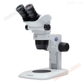
Olympus microscope BX53 fluorescence polarization parameter image quotation
NegotiableUpdate on 03/19
- Model
- Nature of the Manufacturer
- Producers
- Product Category
- Place of Origin
Overview
Olympus Microscope BX53 Fluorescence Polarization Parameter Image Quotation The entire optical path of Olympus Microscope BX53 is designed to optimize fluorescence imaging, using the newly standardized UIS2 component in terms of accuracy and clarity. High quality mirror coating provides excellent transmission and steep cutoff, eliminating almost all stray light from the inner surface for good sensitivity and color separation. Other advanced features further facilitate fluorescence microscopy observation, including a new compound eye lens system that can transmit uniform illumination, as well as automatic magnification and color filter reading functions, simplifying repeated settings.
Product Details
Olympus microscope BX53 fluorescence polarization parameter image quotation
Olympus microscope BX53 fluorescence polarization parameter image quotationShanghai Jintong Instrument National Sales Quotation
The entire optical path is designed to optimize fluorescence imaging, using the UIS2 component that sets new standards for accuracy and clarity. High quality mirror coating provides excellent transmission and steep cutoff, eliminating almost all stray light from the inner surface for good sensitivity and color separation. Other features further facilitate fluorescence microscopy observation, including a new compound eye lens system that can transmit uniform illumination, as well as automatic magnification and color filter reading functions, simplifying repeated settings. In addition, the BX53 semi electric fluorescence microscope enables efficient imaging. Automation can achieve high magnification imaging of large areas and facilitate colorful fluorescence imaging.
High brightness true color LED lighting
The Olympus BX53 microscope has an LED illumination device with a brightness equivalent to 100W halogen lamps, which provides excellent brightness and is very suitable for teaching and polarization applications.
Designed for pathology and cytology
-Clearly view purple, cyan, and pink dyes
-Color temperature *; Don't waste time adjusting color filters
-50000 hours of service life
Maintain the original brightness when changing the magnification
The light intensity manager eliminates the step of adjusting the brightness of the light bulb when changing the magnification, so you can quickly complete observations and reduce eye fatigue.

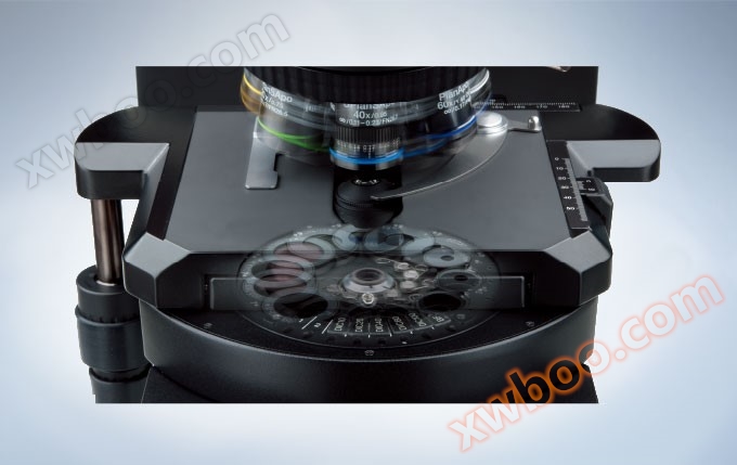
Electric function with rapid amplification factor changes
Easily change the objective lens with an electric objective disc. Place the manual switch near the focusing handle to control the objective lens without taking your eyes off the specimen.
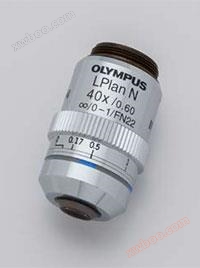
Suitable for cell tissue observation/LPLN40X
Even at a magnification of 40 times, this target is very suitable for imaging clear samples. The LPLN40X is equipped with a correction ring, allowing users to adjust the spherical aberration caused by differences in cover glass thickness to obtain clear images.
Comfortable and easy-to-use stage
Unsupported platform
-Rack mounted, wire drive design, toothless gears help minimize damage
-Mechanical stage, wear-resistant ceramic design
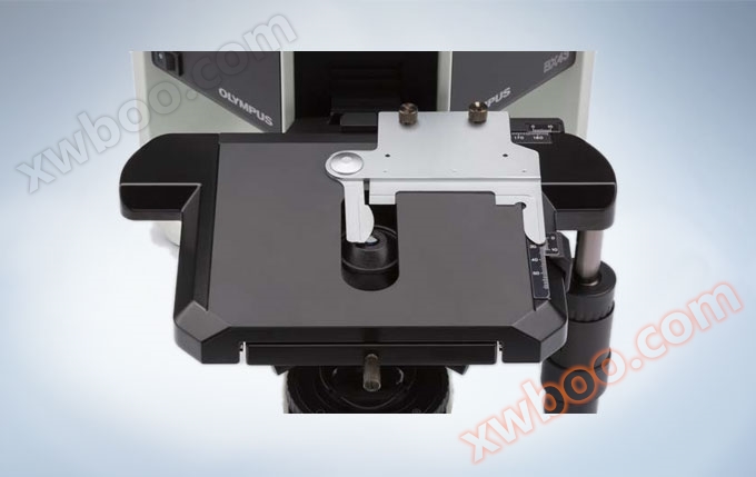
Place your hands on the table
-Place both arms on the table using the stage handle extender
-Install the rubber cap onto the handle and control the stage with small force
Automation can achieve high magnification imaging of large areas and facilitate colorful fluorescence imaging
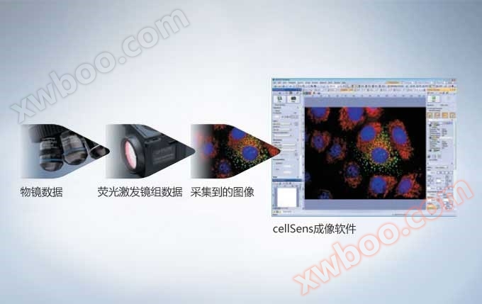
The Olympus BX53 microscope provides greater application flexibility through its semi electric series products and optional automation components, all of which, as well as photography and image acquisition, can be controlled through cellSens software. Therefore, adding these components can provide excellent results for enhancing experiments.
Scalable to meet larger demands
BX3 seriesmicroscopeProvided multiple functions and excellent optical performance for market research. In addition to emphasizing the powerful UIS2 optical system and rigid Y-shaped frame, as well as user-friendly front-end control, it also improves multi-purpose and humanized operation. In addition to emphasizing the powerful UIS2 optical system and rigid Y-shaped frame, as well as user-friendly front-end control, it also improves multi-purpose and humanized operation.
(Source: Chengguan Instrument)
Automatically switch to good contrast settings
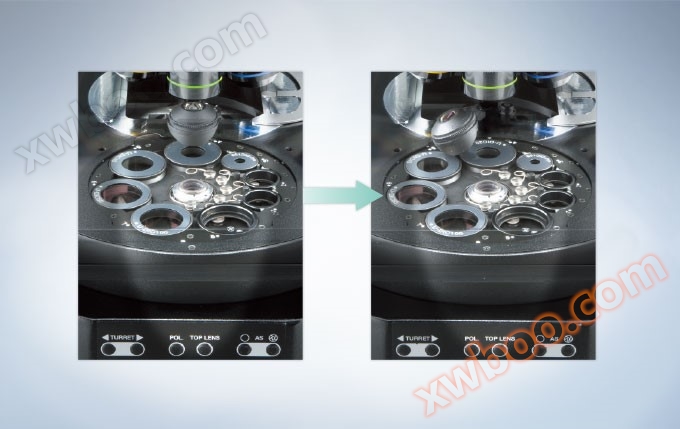
Changing the observation method of transmitted light means ensuring the correct arrangement of ND filter, polarizer/analyzer, and condenser, rather than using the correct onesobjective lensMore important. 8-hole universal spotlight can even control the toplensTo ensure that the entire magnification range can be used. The spotlight ensures easy selection of optical components such as DIC prisms, phase plugins, and polarizing mirrors. To ensure that the illumination light matches the objective lens as much as possible, the aperture stop is automatically set according to the NA of the objective lens in use.
Sensitivity in Fluorescence Imaging
Higher signal-to-noise ratio (S/N) produces fluorescent images with bright colors and dark backgrounds, which is whyOlympusThe reason why it can improve the ability of fluorescence detection and break through the S/N limit. Uniform illumination and detection are achieved through the use of high transmittance objective lenses, excitation lens groups, and integrated compound eye lens systems.(Source: Chengguan Instrument)
Fluorescent Illuminator Using Compound Eye Lens
It is important to ensure uniform illumination in the field of view for the Olympus BX53 microscope. Although the nature of fluorescent lighting makes it a challenging task, Olympus has found a solution based on the concept of fluorescent lighting, which is to integrate a compound eye lens system. As a result of this improvement, users can not only ensure flawless and uniform illumination across the entire wavelength spectrum, but also benefit from simpler light source arrangements.

Compound eye lens surface, magnified image
Integrated flexibility in fluorescent lighting
The Olympus 8-well fluorescence illumination system provides greater flexibility in using multiple fluorescent specimens with its easily replaceable excitation lens group. The need to replace the colorful or FISH excitation lens group has further accelerated the observation operation.

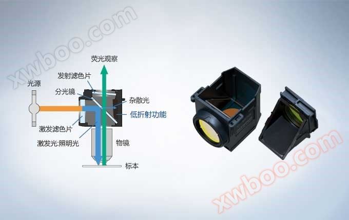
Adopting Olympus coating technology and reducing stray light in the fluorescence excitation mirror group
Optimized UIS2 for fluorescence imagingFluorescence excitation mirror groupSeries products. The high-quality coating used on the excitation lens provides excellent transmittance and steep cutoff slope, while the internal surface eliminates over 99% of stray light, ensuring high sensitivity and excellent color separation. Quickly and easily replace the excitation lens group without the need for tools.(Source: Chengguan Instrument)

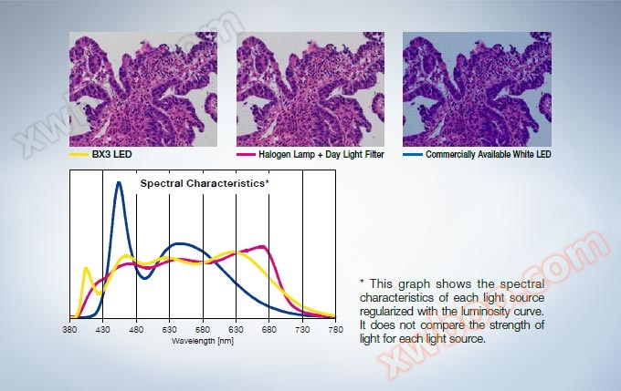
Reduce high transmittance caused by spontaneous fluorescence
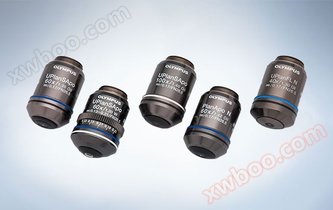
anyoptical microscopeThe core is the quality of the optical system. Olympus Corporation has developed UIS2 optical components to provide excellent optical systems, setting new standards in precision and clarity. Excellent chromatic aberration correction has been achieved for high NA objective lenses, presenting a high resolution that can capture even weak signals. By carefully selecting raw materials for glass slides and using UW multi coating technology, Olympus has reduced the spontaneous fluorescence of the objective lens and greatly improved the S/N ratio. UW multi coating also has high transmittance over a wide wavelength range, ensuring high efficiency in research tasks when using different types of fluorescent dyes.(Source: Chengguan Instrument)
Design a spotlight that reduces back reflection
The electric universal spotlight design can reduce back reflection and spontaneous fluorescence by rotating the top lens, automatically reducing the aperture, and positioning the wheel between two holes during fluorescence imaging.
Low spontaneous fluorescence immersion in oil
The ability to reduce the spontaneous fluorescence typically associated with immersion in oil makes this product * suitable for fluorescence microscopy observation. Noise reduction (spontaneous fluorescence) can increase the signal-to-noise ratio to achieve better fluorescence observation. Reduced the frequency of temporary replacement of fluorescent equipment. It is particularly useful for quantitative observation of single-molecule fluorescence that is easily affected by noise. Anti crystallization allows it to be used for a long time. The refractive index is the same as other Olympus products, ensuring integration into existing microscope observation systems.

Renowned optical performance
When using standard bright field illumination, biological samples do not have inherent contrast, such as color changes, at the microscopic level. Therefore, researchers have adopted a large number of different contrast generation methods. It is mainly divided into two categories: optical contrast method and sampling contrast method. Regardless of the contrast source, the Olympus BX3 series and USI2 optical components perform well, providing sharp and clear images using any contrast method.(Source: Chengguan Instrument)


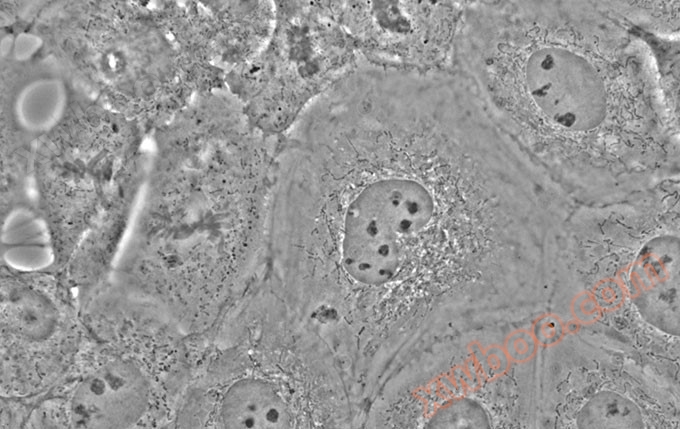
NRK-52E cells (Alexa Fluor 488/Alexa Fluor 546)Polarization observation

Blood island brain during the 12 stages of chicken embryo development (Bodian staining)

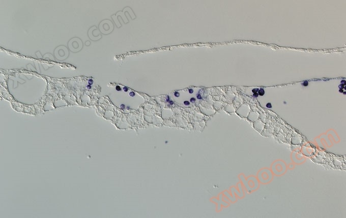
NRK-52E cell water flea
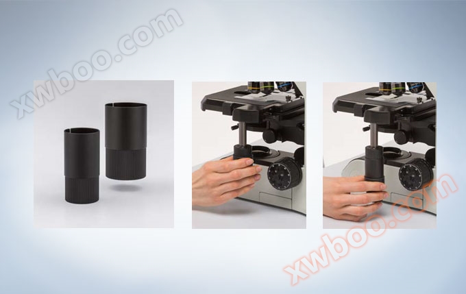
Humanized design for users
Comfortable observation varies from person to person, so it is crucial that the microscope can adapt to every user in all details. Olympus' series of lens barrels provide this capability, allowing for flexible 3D adjustment of tilt angle, extended barrel, and barrel height.
Adjustable height binocular tube
The upright and comfortable posture during microscope operation is not only crucial, but also depends on the user. By adopting humanized barrel style, tilt style, and lift style barrels, the tilt angle can be adjusted, the barrel and barrel height can be extended, and flexible 3D assembly can be achieved. Thus adjusting the microscope to accurately adapt to the user, rather than the opposite.

Binocular tube that meets every need
We provide a variety of tilted binocular barrels to meet various needs. A model that generates traditioninvertObserving the image, while other models produce movements in the same direction as the specimenregularityImage; This makes it easier to locate specific areas in the specimen.

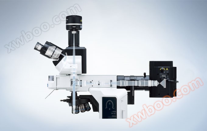
Intuitive control solution for meetings and imaging
Like quality, true flexibility is also a characteristic that must be designed into the instruments at each stage. Based on this goal, Olympus Corporation has developedUpright research microscopeThe BX3 series. BX53 is a representative product of high-performance imaging systems; Provides a wide range of optical, semi electric components and software options, as well as continuous adaptability during use.
Tilted three eye tube for good posture
Each user has specific posture and positioning requirements, therefore, the ability to adjust microscope controls, coupled with the activation of personalized workspaces, helps provide a humanized environment suitable for long-term microscope use without causing posture injuries or repeated strains. To achieve greater system flexibility and user comfort, the tilted humanized three lens barrel provides excellent eyepiece height adjustment and pupillary distance control. The light path slider can be installed on both sides of the barrel, allowing users to fully control the microscope and confirm that it can be adjusted to suit their posture.(Source: Chengguan Instrument)

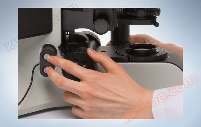
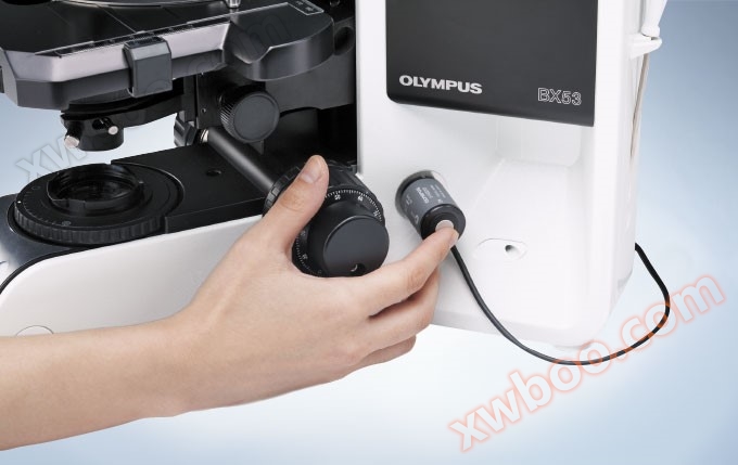
Manual switch for image acquisition
The Olympus BX53 remote exposure knob can be installed on any side of the microscope, and image acquisition can be performed by touching the button, without the need for users to turn around from the microscope to view the display and use a mouse. This is not only more effective, but also more user-friendly for users.
Microscopic observation data saved using encoding devices
The BX3 series comes with an optional manual optionobjective lensThe converter and encoder mirror group converter module enables users to automatically record and share microscope magnification and setting information, facilitating post-processing of imaging. This reading function is for OlympusCellSens softwareThe package automatically provides correct metadata to ensure that there are no errors or scale errors when recording images.
Design a system that meets expansion needs
Olympus Research MicroscopeBX53Observation does not only include microscopes with imaging capabilities, as each examination requires specific settings. Therefore, each system must not only be highly flexible, but also adept at developing and handling a large number of solutions. The Olympus BX3 series microscope is such a device that achieves excellent modularization of hardware and software, embedded in a flexible imaging system environment, ensuring that researchers can always maintain control of any task.(Source: Chengguan Instrument)
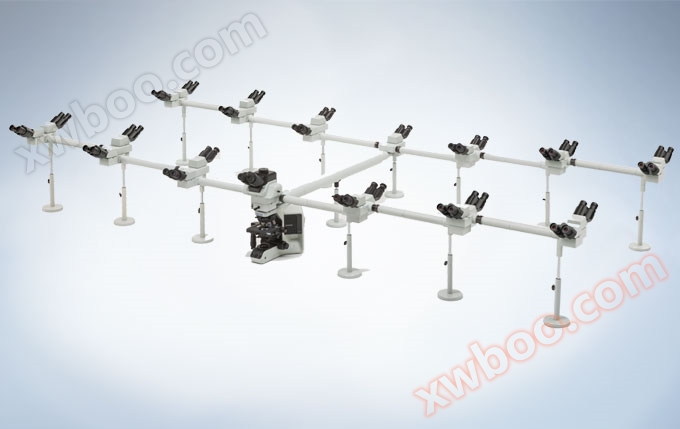
Group observation system
In addition to user-friendly binocular and trinocular tubes, Olympus also provides customizable two person and multi person observation accessories for laboratory observation. These systems also have immeasurable effects on clinical observation, teaching, and guidance, and it would be more effective if the entire team could observe and discuss specimens through their respective eyepieces. There can be two to ten participants, or even more people participating.
Digital imaging that meets different needs
The multi-purpose BX53 system is not just a microscope, it can be easily assembled into a complex imaging system to meet any application needs. From models used for cutting-edge research work to excellent single machine microscopes used for conferences. Our entire seriesdigital cameraandcellSensImaging SoftwareEnsure high pixel color fidelity collection of tissue staining slides during all clinical diagnoses.
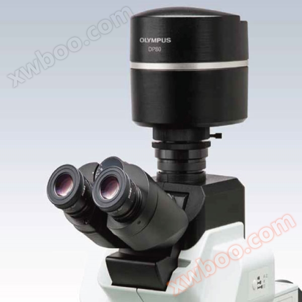
DP80 digital camera
DP80 is a * camera that combines color and monochrome sensors in the same chassis, providing high-resolution bright field imaging and sensitive photon detection of fluorescent dyes. Using pixel shift technology to achieve a recording image size of 4080 × 3072, this cellSens control software with multiple functions and high resolution equivalent to 12.5 million pixels can achieve fast automatic switching between chips without manually switching microscope light paths. In addition, DP80 is able to overlay images from two sensors with precise pixel to pixel correspondence, providing effective and accurate combined color and fluorescence imaging in research and clinical environments.
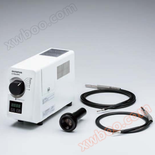
Optical coupling illumination fluorescent light source U-HGLGPS
The 130W U-HGLGPS optical coupling illumination system is an ideal stable light source for fluorescence microscopy observation.OlympusOptical coupling illumination fluorescent light sourceU-HGLGPSThe long-lasting and powerful pre aligned light source reduces operating costs, is easy to install, and has excellent long-term and short-term stability. Its six level aperture can effectively control the light intensity and implement simple, graded intensity adjustment. The coupling of optical fibers reduces thermal effects and vibrations at the microscope frame, ensuring good experimental conditions.
imaging system
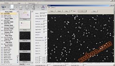
Sperm quality analysis software
The MMC HST sperm quality analysis system is a well-known computer-aided sperm analysis system (CASA). The use of advanced computer technology for automated quantitative detection and analysis of sperm density, vitality, viability, and movement trajectory characteristics has been published in various renowned academic journals. The entire HST system consists ofmicroscopicConsisting of amplification devices, digital quick capture devices, software packages, and workstations. This system is used for clinical semen examination and can greatly improve the level of clinical testing. It is widely used in urology, urology, obstetrics and gynecology (in vitro fertilization), family planning, and eugenics research in hospitals. This system is used in certain professional fields, such asbiologyResearch and animal studies (such as pigs, cows, sheep, pandas, fish, etc.) also have wide application value. The fully automated analysis software package MMC is used for semen quality analysis based on the recommended parameters of the World Health Organization (WHO). This software package is based on our professional database system and has specialized sperm analysis procedures.More details
Fluorescence in situ hybridization FISH software
Imstar, a French company, was founded in 1985 and is headquartered in the charming city of Paris. For 28 years, we have been committed to the field of fully automated and intelligent large-scale injection scanning image analysis, and have always been in a technological position in this research field. The fields covered by Imstar's products include cellular pathology, cytogenetics, genetic toxicology, drug safety evaluation, environmental monitoring, life science research, and drug development. The software includes chromosome karyotyping analysis, fluorescence in situ hybridization (FISH) analysis, HER/2, fluorescence point counting, and multi-layer scanning depth of field extended confocal microscopy.More details

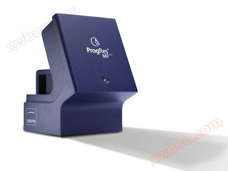
Jena microscope digital camera
Jenoptik ProgRes ® The MFcool microscope camera has accelerated the work speed of the laboratory. This is thanks to its high image refresh rate. This camera is equipped with a sensitive monochrome CCD sensor and is specifically designed for scientific research applications. Its high sensitivity means that clear and accurate image results can be obtained even under low light conditions.More details
Similar Product Recommend