-
Phone
1376488515213764885152
-
Address
Building 6, No. 1150 Lanfeng Road, Fengxian District, 4597
Product Categories
Shanghai Jintong Instrument Co., Ltd
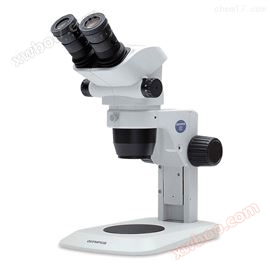
SZX16 Olympus research grade clinical stereomicroscope
NegotiableUpdate on 03/19
- Model
- Nature of the Manufacturer
- Producers
- Product Category
- Place of Origin
Overview
SZX16 is designed for advanced research and is suitable for oblique and bright field illumination. Its numerical aperture (NA) of 0.3 produces a resolution of 900 pairs of lines per millimeter, enabling effective acquisition of more sample information. The advanced stereo device with a super large zoom ratio includes an observation tube, a zoom lens body, and an objective lens, achieving * color correction and greatly reducing the color difference blur throughout the entire magnification range. Olympus research grade clinical stereomicroscope.
Product Details
Olympus research grade clinical stereomicroscope
(1) Excellent optical performance
The SZX16 optical components, including the observation tube, zoom lens body, and objective lens, have achieved * color correction, reducing the chromatic aberration blur of the entire magnification range to a certain extent. The SDF (Super Depth of Focus) objective lens can provide clear and high contrast images of any specimen. An additional 0.3NA resulted in a resolution of 900 pairs of lines per millimeter, enabling clear observation of cells and cell structures under a microscope. This resolution and magnification make the work more efficient, accurate, and can display more information about the sample.
(2) 16.4:1 zoom ratio
The SZX16 has a magnification zoom ratio of 16.4:1, which can be achieved with a 1x objective for 7x to 115x magnification, and with a 2x objective for up to 230x magnification. This zoom range makes it an ideal choice for life science applications that require low magnification macroscopic observation (for dissection and specimen processing) and ultra sharp high magnification observation (for close range observation of cell structures).
(3) Dual objective lens with zoom ranging from 3.5x to 230x
The use of a focal length objective lens and a dual hole position objective lens converter enables fast and easy switching between objectives, with almost no need for refocusing. Therefore, after using 0.5x and 2.0x objective lenses, the continuous magnification range ranges from 3.5x to 230x. This indicates an effective zoom ratio of 65.7:1.
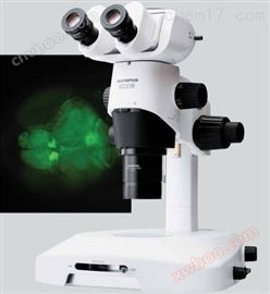
(4) Advanced fluorescence observation
SZX16 has precision manufactured optical components and high NA values, making it an ideal fluorescence microscope for observing details from entire tissues to individual cell nuclei. Advanced glass materials with low spontaneous fluorescence performance and new surface coating technologies greatly increase the transmittance of light, thereby improving the clarity of images. In addition, these performances also provide a high S/N ratio and high fluorescence signal intensity. The five hole color filter turntable facilitates comprehensive control of excitation light and enables flexible illumination from low magnification to high-resolution imaging.
(5) SZX16 filter disk for fluorescence observation
Left: zebrafish embryos expressing GFP, right: zebrafish embryos expressing GFP (28-30 hours). The image data is provided by the Developmental Gene Regulation Laboratory of RIKEN Institute of Brain Science
(6) High resolution with large depth of focus
The SDF (Super Depth of Focus) objective lens can provide clear high contrast images of any specimen. SZX16 has six SDF objective lenses, four of which are collimated flat field apochromatic lenses: 0.5x, 1.0x, 1.6x, and 2.0x. The 0.3x half achromatic objective and 0.8x flat field achromatic objective have an extended working distance.
(7) SDF Objective Lens Series
Optimize for samples in the culture medium
The 2.0x SDF objective of SZX16 has a good NA of 0.3. This flat field apochromatic objective is more suitable for tissues and cells in culture medium. Its correction ring can fine tune the lens to correct mismatches caused by the different refractive indices of the container and culture medium. This helps achieve possible resolution when using Petri dishes and other culture dishes.
(8) Excellent fluorescence performance
The excellent NA value characteristics of SDF objectives greatly improve fluorescence sensitivity. In addition, the excitation light path of the newly designed approximate vertical reflection light illuminating device is independent of the observation path, which can effectively improve the efficiency of excitation light. These features provide brightness far superior to traditional stereomicroscopes in fluorescence observation at all magnifications. Even in reflective fluorescence observation, transmitted light observation can be used to verify the contour of the specimen.
Practical design for advanced research
(9) Electric solutions
The SZX16 electric focusing drive uses a manual switch, simplifying focusing and zooming. And make digital recording with depth of field extension (EFI) function efficient and automated. It is even possible to create pseudo 3D images, closing the gaps between the recorded files and the images observed through the optical path of a stereo microscope. For the convenience of automatic inspection, Olympus cellSens software provides all the tools needed for simple 2D measurements to complex phase analysis in an easy-to-use environment.
(10) Spacious space for control
Any type of manipulation under a microscope requires ample space between the top of the sample and the bottom of the objective lens for tool use. The objective lens used in SZX16 has a long working distance and can be operated manually and automatically with injection tools. In addition, due to the gradual thinning of the front end of the 1.6x and 2x high-power objective lenses, the effective movement range of the controller is greatly increased.
(11) ComfortView eyepiece for reducing eye fatigue
The advanced ComfortView eyepiece provides a larger range of eye movement for observing 3D images, making short-term and long-term use more comfortable. This also makes it easier to form stereoscopic images and reduces the occurrence of eye fatigue. And the optical coating technology also presents true color images with exquisite details.
Comfortable eyepiece for observation
(12) Humanized observation tube
The continuously adjustable tilt binocular tube (5 to 45 degrees) allows the operator to quickly find the appropriate viewpoint position, reducing the risk of back strain and muscle tension to a certain extent, and improving efficiency and quality in daily applications.
Adjustable convergence angle of the lens barrel, tilted observation tube
Multiple attachments
(13) Lighting base selection
SZX2-ILLT
Ultra slim transmission light solution for normal bright field, contrast enhanced bright field, dark field, and oblique illumination. The bright LED base is only 41mm high, half the height of a normal base, but provides amazing flexibility, humanization, and durability for experienced and first-time users.
SZX2-ILLK
Transmitting light bracket for bright field and oblique illumination.
Equipped with a built-in 6V/30W halogen lamp, it provides contrast enhanced images for transparent specimens.
SZX2-ILLD
Transmitting light support for both bright and dark fields. Equipped with a built-in 6V/30W halogen lamp, it can switch between bright and dark field lighting; Reduced background noise, improved contrast, and able to distinguish even subtle details.
SZX2-ILLB
High level transmission light bracket for bright field and oblique illumination.
The selection of high and low contrast provides clear and effective contrast and illumination.
It is easy to adjust the light intensity and color temperature on 6V/30W halogen lamps.
When used in combination with a high-power objective lens, it can observe extremely small details on high contrast structures; Especially effective against C. elegans, oocytes, and embryos.
(14) Digital imaging and recording
The SZX16 can also be equipped with a three lens barrel and an Olympus DP series digital camera for high-resolution imaging and research recording. The DP series cameras provide high sensitivity for a wide range of applications, including high-sensitivity fluorescence imaging, and can be easily controlled using Olympus cellSens software.
Microscope with three lens barrel and DP series digital camera related to Olympus cellSens software
(15) Technical parameters
Observation method |
Fluorescence (blue/green excitation) |
? |
||
Fluorescence (UV excitation) |
? |
|||
Simple polarization |
? |
|||
bright field |
? |
|||
dark field |
? |
|||
zoom |
Zoom magnification |
16.4 |
||
Magnification indicator |
0.7, 0.8, 1, 1.25, 1.6, 2, 2.5, 3.2, 4, 5, 6.3, 8, 10, 11.5 |
|||
electric |
? |
|||
AS |
·Built in |
|||
feature |
Variable magnification amplification system with parallel optical axes |
|||
Optical components |
Galilean optical system |
? |
||
Illuminator |
fluorescent illuminator |
mercury lamp |
· ? |
|
xenon lamp |
· ? |
|||
Optical lighting |
· ? |
|||
focus |
Focusing device |
Coarse/fine adjustment focus |
? |
|
electric |
· ? |
|||
load capacity |
·Manual: 0-10.0kg/2.7-15.0kg/8.0-25.0kg ·Electric: 0.0-23.0kg |
|||
Handle stroke |
·Manual: 80mm/120mm ·Electric: 78 mm |
|||
Rough adjustment handle stroke for each rotation |
·Manual: 36.8 mm ·Electric: 2.7 mm |
|||
Fine tuning handle stroke for each rotation |
·Manual: 0.77 mm ·Electric: 0.27 mm |
|||
Electric zoom |
? |
|||
Nosepiece |
manual |
standard form |
? |
|
barrel |
Wide Field of View (FN22) |
trinocular tube |
? |
|
Tilted three lens tube |
? |
|||
Tilt angle of the lens barrel |
· 5 - 45° · 30° |
|||
Three lens tube optical path (camera: visible to the naked eye) |
· 50 % : 50 %, 0 % : 100 % · 0 % : 100 %, 100 % : 0 % |
|||
Interpupillary distance adjustment |
· 51 - 76 mm · 52 - 76 mm |
|||
frame |
Standard rack |
? |
||
Optional base and stand |
Thin LED Transmitting Light Illumination Base |
? |
||
High level transmitted light base |
? |
|||
Bright/dark field transmitted light illumination base |
? |
|||
Large scale scaffolding |
? |
|||
Wanxiang energy platform |
? |
|||
Overall dimensions |
285 (W) x 335 (D) x 403 (H) mm (standard configuration) |
|||
operating environment |
For indoor use |
ambient temperature |
0 - 40 oC (32 - 104 oF) |
|
relative humidity |
30 - 90 % |
|||
eyepiece |
· WHSZ10X-H FN 22* · WHSZ15X-H FN 16* · WHSZ20X-H FN 12.5* · WHSZ30X-H FN 7* ·*With diopter adjustment and marking |
|||
objective lens |
SDFPLFL0.3X, WD 141 mm, NA 0.045 |
|||
fluorescent |
Nearly vertical fluorescent lighting fixture |
|||
Olympus research grade clinical stereomicroscope
Similar Product Recommend