-
E-mail
sales-sci.cn@horiba.com
-
Phone
13681872955
-
Address
Unit D, 1st Floor, Building A, Lianqiang International Plaza, 1068 Tianshan West Road, Shanghai
Product Categories
HORIBA (China)

AFM optical workstation
NegotiableUpdate on 01/04
- Model
- Nature of the Manufacturer
- Producers
- Product Category
- Place of Origin
Overview
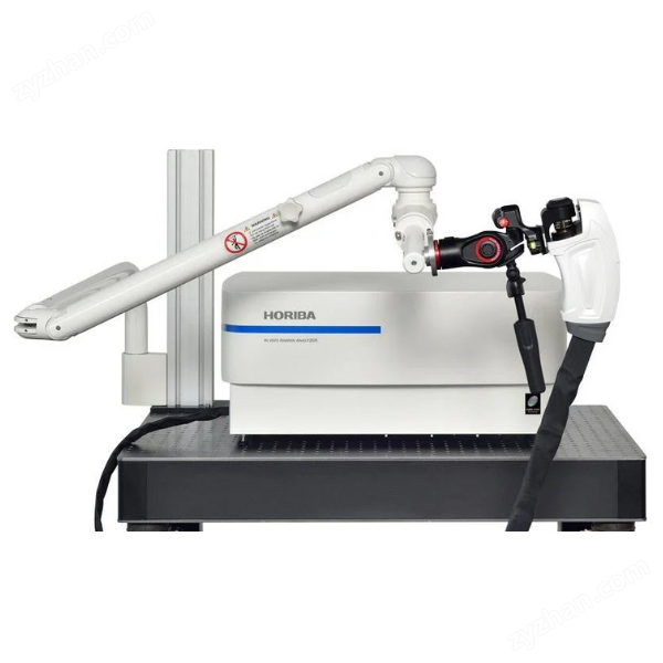
The OmegaScope AFM optical workstation is a cutting-edge solution for coupling optics and AFM high-resolution, multidisciplinary research, providing the latest avenues for spectroscopy and photonics researchers. It provides a reflective configuration with direct top and lateral optical path coupling. OmegaScope has built a flexible platform for coupling imaging modes related to high spatial resolution spectroscopy (Raman, photoluminescence, fluorescence) and AFM.
Product Details
OmegaScopeAFM optical workstation
OmegaScope is a cutting-edge solution for coupling optics and AFM high-resolution, multidisciplinary research, providing the latest avenues for spectroscopy and photonics researchers. It provides a reflective configuration with direct top and lateral optical path coupling. OmegaScope has built a flexible platform for coupling imaging modes related to high spatial resolution spectroscopy (Raman, photoluminescence, fluorescence) and AFM.
Raman laser and AFM feedback laser have no interference with each other
The 1300nm atomic force microscope feedback laser and commonly used ultraviolet, visible, and near-infrared Raman lasers (364-830nm) have no mutual interference and eliminate any parasitic effects on visible light sensitive biological and photovoltaic samples.
Raman laser direct to cantilever
The OmegaScope system separates AFM and optical channels. This independence does not limit the wavelength required for Raman laser, simplifying the adjustment of the entire system compared to AFM laser through the same high aperture objective lens as Raman excitation laser. Users can easily refocus high NA objectives without any additional adjustments to the AFM laser. The design of OmegaScope provides higher stability for atomic force microscopy and reduces sensitivity to vibration and noise.
Simple, fast, and repeatable cantilever adjustment
The design adopts a fixed AFM laser to excite the laser and adjust the tip of the cantilever beam, which is simple and fast. In addition, if a new cantilever of the same type is installed, it is easy to find and scan the same area on the surface of the sample (within a repeatability range of a few micrometers) without any additional search.
Automatic AFM registration system adjustment
The SmartSPM scanning probe microscope is the core configuration of the reflective mechanism in the OmegaScope system, and it is also the first scanning probe microscope to use laser cantilever photodiode fully automatic collimation from design to coupling with HORIBA spectrometer.
quick scan
Adopting the industry's highest resonant frequency scanner (XY>7kHz and Z>15kHz), the optimized scanner control algorithm can easily achieve fast scanning.
Vibration stability, acoustic stability, high-frequency fast scanner
Fast response time, low drift, and value traceability. Adopting the industry's best flat closed-loop scanner, with a scanning range of 100 μ m x 100 μ m x 15 μ m, a single scanner can achieve large-scale measurement to true molecular resolution imaging. Thanks to the high mechanical stiffness of the scanner and the entire atomic force microscope, OmegaScope's outstanding performance can be guaranteed even without active shock absorption protection. These features also allow for the implementation of special and more complex scanning algorithms, such as Top mode. In this mode, the needle tip is lifted above the sample surface between scanning points. At each scanning point, the probe will return to the surface of the sample. The scanning signal is measured immediately after the needle tip oscillation amplitude reaches the set threshold. It can avoid any interaction of lateral forces, such as ensuring the safety of TERS probes, while maintaining a scanning rate of up to 1Hz.
Easy replacement of samples
The OmegaScope AFM platform design allows for sample replacement without removing the FM head and cantilever bracket. It greatly improves the reliability of the experiment and avoids errors that operators may make during such routine processes.
Top and side lighting
Both the top and lateral optical channels can enter the needle tip sample area, fully utilizing the spectral imaging capabilities of infrared, visible light, and ultraviolet high NA achromatic objective lenses (top objective lens: up to 0.7NA; lateral objective lens: up to 0.7NA) and atomic force microscopy related technologies, enabling confocal detection of optical signals on the sample surface within a wide spectral range and minimum excitation laser spot area. The success of the lateral optical path in TERS and TEPL experiments is attributed to the rationality of the OmegaScope system design, which provides a more important axial component of the electromagnetic field and effectively excites plasmon resonance in the needle tip sample junction.
Top and lateral objective scanner
In order to align the AFM tip with the Raman laser beam, a flat closed-loop XYZ objective scanner can be installed at the top, side, and bottom. In addition, this solution provides the highest possible resolution, long-term stability, and alignment automation, coupled with a wider spectral range and minimal optical components in the input/output system, greatly reducing the loss of useful optical signals.
Built in DFM measurement controlled by phase-locked loop
The Dynamic Force Microscopy (DFM) mode is a standard configuration of the OmegaScope system. A frequency modulation (FM) detector suitable for this mode was designed using the built-in phase-locked loop (PLL) circuit of the controller. The use of DFM can reliably maintain minimal needle sample interaction (i.e. imaging within the attractive region), which is crucial for successful TERS and scanning near-field optical microscopy (SNOM) experiments.
STM、 Conductive AFM and SNOM options
Along with spectral measurement, OmegaScope can be equipped with a module that can measure local currents in three linear ranges (1nA, 100nA, and 10uA) in AFM or STM. These ranges can be switched in the software, and the required bandwidth for each range can be selected from 100Hz to 7kHz. The noise level of the 60fA conductive module has set a new standard for conductivity measurement in the field of optoelectronics within the measurement range of 1na and 1300nm atomic force microscope lasers.
In addition to the special flexibility of the OmegaScope platform, SNOM based on tuning fork feedback design is also provided. In addition to standard SNOM experiments, you can also follow classical nanophotonics, especially aperture free SNOM, using a metal needle tip illuminated by femtosecond laser pulses with appropriate polarization for near-field fluorescence imaging.
SmartSPM scanner and base
Closed loop flatbed scanner: 100 µ m x 100 µ m x 15 µ m (± 10%)
Scanner nonlinearity: XY ≤ 0.05%; Z≤0.05 %
Noise level: XY ≤ 0.1 nm RMS (200 Hz bandwidth, capacitive sensor turned on);
XY ≤ 0.02 nm RMS (100 Hz bandwidth, capacitive sensor turned off);
Z<0.04 nm RMS (1000 Hz bandwidth, capacitive sensor on)
High frequency scanner: XY ≥ 7 kHz; Z≥ 15 kHz
X. Y, Z automatic approach: XYZ digital closed-loop control, Z-direction motor approach distance of 18mm
Sample size: 40 mm x 50 mm x 15 mm
Sample positioning: Automatic sample stage range: 5 mm x 5 mm
Positioning accuracy: 1 µ m
AFM test head HE002
Laser wavelength: 1300nm
Laser has no effect on biological samples;
Laser has no effect on photoelectric measurement;
System noise:<0.1 nm
Fully electric: 4-step motor for automatic alignment of cantilever and photodiode;
Probe channel: provides a free channel for external operations and probes;
Top and lateral simultaneous optical path channels: equipped with flat achromatic objective lens, can simultaneously achieve up to 100x, NA0.7 lateral objective lens and 10x, NA0.28 top objective lens;
SPM measurement mode
Standard modes: contact mode, semi contact mode, non-contact mode, phase imaging mode, lateral force mode (LFM), force modulation mode, magnetic force microscopy mode (MFM), Kelvin probe mode (surface potential, SKM, KPFM), scanning capacitance mode, electrostatic force microscopy mode (EFM), force curve measurement, piezoelectric response mode (PFM), nanoetching, nanomanipulation
Upgrade modes: solution environment contact mode, solution environment semi contact mode, conductive force microscope mode, STM mode, photocurrent imaging mode, volt ampere characteristic curve measurement
Synchronous Raman SPM measurement mode
Aerial contact atomic force microscope;
Liquid contact atomic force microscope (optional);
Half contact atomic force microscope in air;
Liquid semi contact atomic force microscope (optional);
Dynamic Force Microscopy (DFM, FM-AFM);
Dissipative force microscope;
True non-contact atomic force microscope;
Phase imaging;
Lateral force microscope;
Force modulation;
Conductive atomic force microscope (optional);
Single channel Kelvin probe;
Piezoelectric response force microscope;
STM (optional);
Photocurrent imaging (optional);
Shear force microscope with tuning fork (ShFM) (optional);
Normal force microscope with tuning fork (optional).
Spectral mode
Confocal Raman, Fluorescence, and photoluminescence spectroscopy and imaging
Needle tip enhanced Raman spectroscopy (AFM, STM, etc.)
Needle tip enhanced fluorescence
Near Field Optical Microscopy and Spectroscopy (NSOM/SNOM)
Current range: 100fA~10 µ A; automatic switching of three ranges (1 nA, 100 nA, and 10 µ A)
Conductive AFM (optional)
Current range: 100fA~10 µ A; automatic switching of three ranges (1 nA, 100 nA, and 10 µ A)
Optical path coupling channel
The top and side can use achromatic lenses simultaneously: up to 100X, NA0.7 lenses can be used from the top or side; Can use both 20x and 100x simultaneously
Long term spectral laser stable alignment closed-loop piezoelectric objective scanner: 20 µ m x 20 µ m x 15 µ m; resolution: 1nm
OmegaScopeAFM optical workstation